. 2017 Sep 1;144(17):3177-3188.
doi: 10.1242/dev.149112. Epub 2017 Jul 13.

Cellular heterogeneity in the ureteric progenitor niche and distinct profiles of branching morphogenesis in organ development

Affiliations

Cellular heterogeneity in the ureteric progenitor niche and distinct profiles of branching morphogenesis in organ development

Elisabeth A Rutledge et al. Development. .

Abstract

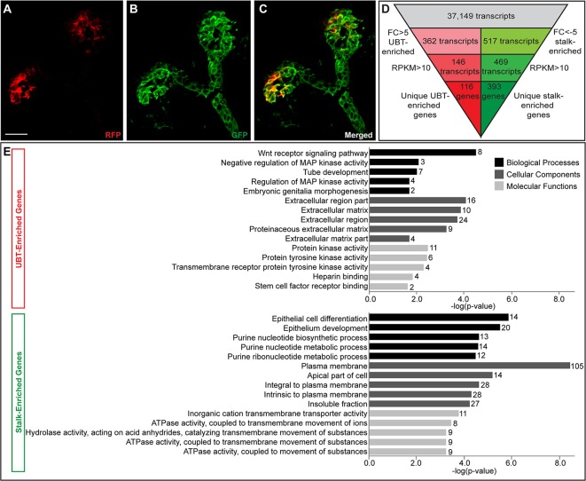
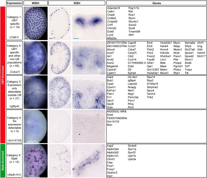
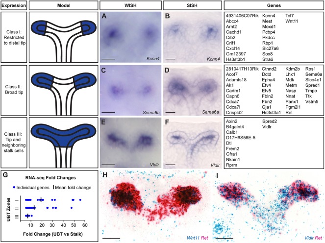
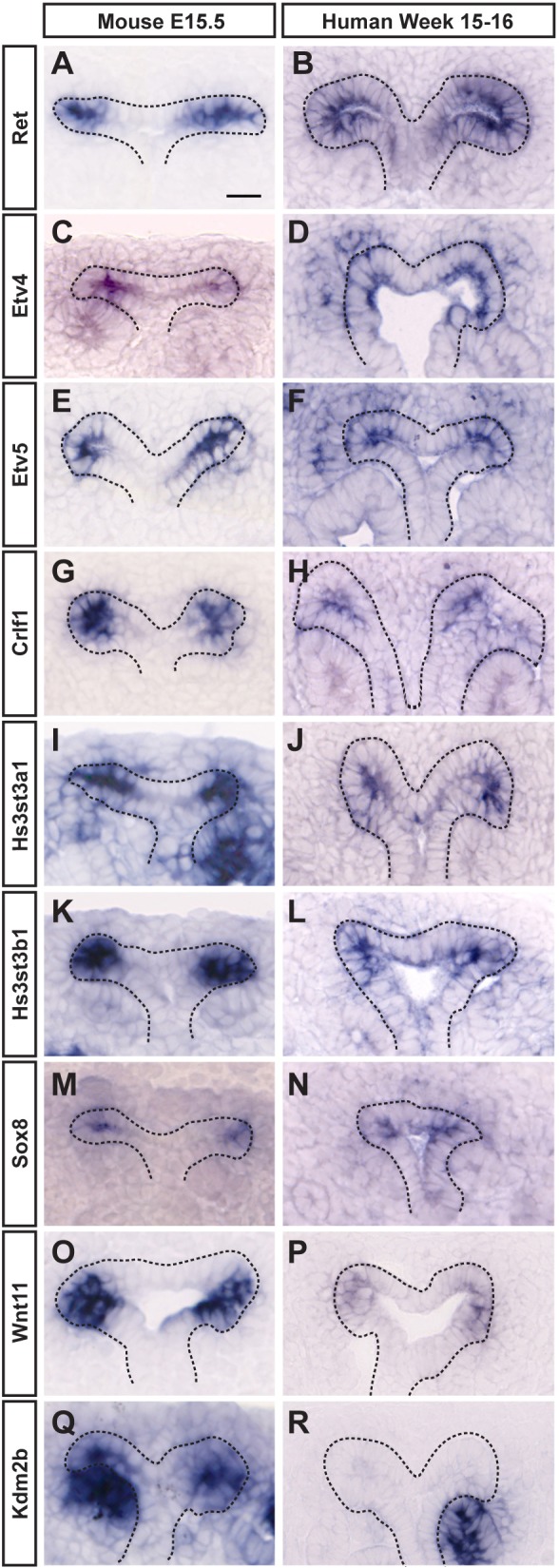
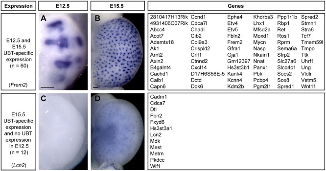
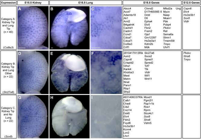
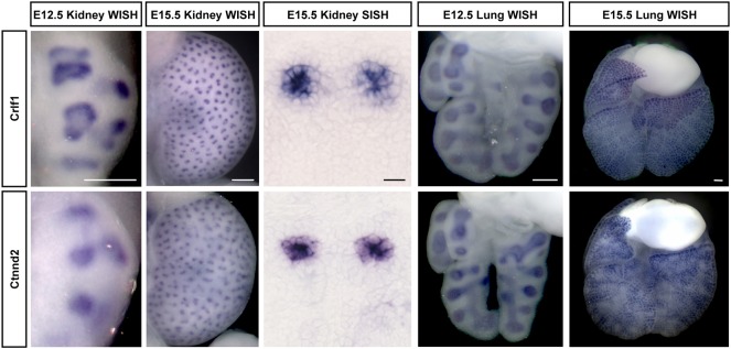
Branching morphogenesis creates arborized epithelial networks. In the mammalian kidney, an epithelial progenitor pool at ureteric branch tips (UBTs) creates the urine-transporting collecting system. Using region-specific mouse reporter strains, we performed an RNA-seq screen, identifying tip- and stalk-enriched gene sets in the developing collecting duct system. Detailed in situ hybridization studies of tip-enriched predictions identified UBT-enriched gene sets conserved between the mouse and human kidney. Comparative spatial analysis of their UBT niche expression highlighted distinct patterns of gene expression revealing novel molecular heterogeneity within the UBT progenitor population. To identify kidney-specific and shared programs of branching morphogenesis, comparative expression studies on the developing mouse lung were combined with in silico analysis of the developing mouse salivary gland. These studies highlight a shared gene set with multi-organ tip enrichment and a gene set specific to UBTs. This comprehensive analysis extends our current understanding of the ureteric branch tip niche.

Keywords: Branching morphogenesis; RNA-seq; Tip progenitor; Ureteric bud.


Conflict of interest statement

Competing interestsThe authors declare no competing or financial interests.

Figures

Fig. 1.
Fig. 1.
Identification of UBT- and stalk-enriched genes from RNA-sequencing data set. (A-C) Immunofluorescence of a Wnt11RFP;Hoxb7GFP E15.5 ureteric bud used to acquire UBT and stalk cell populations for RNA sequencing. (A) RFP-positive cells restricted to the UBTs. (B) GFP-positive cells labeling the entire ureteric epithelium. (C) Merged image displaying the UBT cells (RFP+, GFP+) and stalk cells (GFP+). (D) Filtering strategy to narrow down to a stringent list of UBT- and stalk-enriched genes for further analysis. (E) Gene ontology (GO) term analysis of UBT-enriched and stalk-enriched gene lists (number of genes for each term are listed at the end of the bar). Scale bar: 20 μm.
Fig. 2.
Fig. 2.
Whole-mount and section in situ hybridization expression pattern analysis of UBT-enriched and stalk-enriched genes identified from the RNA-seq dataset on Swiss Webster E15.5 kidneys. Expression patterns shown are example genes for each category as indicated on the left. Genes listed on the right have the expression pattern described in the categories on the left. Leprel2, also known as P3h3. *In addition to ureteric tip-restricted expression in the ureteric epithelium, Wnt11 is activated in interstitial mesenchyme cells, coincident with medullary remodeling from E15.5 (Yu et al., 2009). Scale bars: 100 μm (black); 20 μm (blue).
Fig. 3.
Fig. 3.
Classification of UBT expression from whole-mount and section in situ hybridization within E15.5 kidneys highlighting three distinct zones. (A,B) Kcnn4 is restricted to the very distal ends of the UBT. (C,D) Sema6a is expressed widely throughout the tip domain. (E,F) Vldlr is expressed throughout the UBT domain and weakly into the near adjacent stalk regions. Schematics illustrating the expression domains for each class are shown on the left. Expression patterns shown in A-F are example genes for each class as indicated. Genes categorized within each class are listed on the right. (G) Distribution of RNA-seq fold changes for each zone. (H) Expression pattern of Wnt11 (Class I) and Ret (Class II). (I) Expression pattern of Ret (Class II) and Vldlr (Class III). Scale bar: 20 μm.
Fig. 4.
Fig. 4.
SISH comparison of UBT expression between mouse (E15.5) and human (week 15-16) kidneys. (A-N) Ret, Etv4, Etv5, Crlf1, Hs3st3a1, Hs3st3b1 and Sox8 show strong UBT expression in both species. (O,P) Wnt11 has strong UBT expression in the mouse and much weaker UBT expression in the human kidney. (Q,R) Kdm2b shows UBT expression only in the mouse kidney. Note that expression in the distal portion of the renal vesicle is conserved between the species. Scale bar: 20 μm.
Fig. 5.
Fig. 5.
WISH UBT expression in Swiss Webster kidneys at E12.5 and E15.5. (A,B) Frem2 has UBT-specific expression in the kidney at both E12.5 and E15.5. (C,D) Lcn2 only shows UBT expression in the E15.5 kidney. Scale bars: 100 μm.
Fig. 6.
Fig. 6.
Expression pattern analysis of tip-enriched genes in Swiss Webster E15.5 kidneys and lungs. (A-C) Col9a3 shows tip expression in both kidneys and lungs at E15.5. (D-F) Slc27a6 is UBT specific in the kidney and is expressed outside of the tip in the lung. (G-I) Sox8 is expressed within the kidney UBT and is absent from the lung. Expression patterns shown are example genes for each category as indicated on the left. Italicized genes have a different tip expression pattern at E12.5 compared with E15.5, which are categorized on the right under ‘E12.5 Genes’. Scale bars: 200 μm.
Fig. 7.
Fig. 7.
Examples of kidney and lung expression of two UBT-specific genes characterized by in situ hybridization at E12.5 and E15.5. Scale bars: 200 μm (white); 20 μm (black).

References

    1. Affolter M. and Caussinus E. (2008). Tracheal branching morphogenesis in Drosophila: new insights into cell behaviour and organ architecture. Development 135, 2055-2064. 10.1242/dev.014498 - DOI - PubMed
    1. Arman E., Haffner-Krausz R., Gorivodsky M. and Lonai P. (1999). Fgfr2 is required for limb outgrowth and lung-branching morphogenesis. Proc. Natl. Acad. Sci. USA 96, 11895-11899. 10.1073/pnas.96.21.11895 - DOI - PMC - PubMed
    1. Belle M., Parray A., Belle M., Chédotal A. and Nguyen-Ba-Charvet K. T. (2016). PlexinA2 and Sema6A are required for retinal progenitor cell migration. Dev. Growth Differ. 58, 492-502. 10.1111/dgd.12298 - DOI - PubMed
    1. Blomqvist S. R., Vidarsson H., Fitzgerald S., Johansson B. R., Ollerstam A., Brown R., Persson A. E. G., Bergström G. G. and Enerbäck S. (2004). Distal renal tubular acidosis in mice that lack the forkhead transcription factor Foxi1. J. Clin. Invest. 113, 1560-1570. 10.1172/JCI20665 - DOI - PMC - PubMed
    1. Bock H. H. and May P. (2016). Canonical and non-canonical reelin signaling. Front. Cell Neurosci. 10, 166 10.3389/fncel.2016.00166 - DOI - PMC - PubMed

Publication types