. 2017 Jul 13;7(1):5298.
doi: 10.1038/s41598-017-05647-9.

Connexin 50 Functions as an Adhesive Molecule and Promotes Lens Cell Differentiation

Affiliations

Connexin 50 Functions as an Adhesive Molecule and Promotes Lens Cell Differentiation

Zhengping Hu et al. Sci Rep. .

Abstract

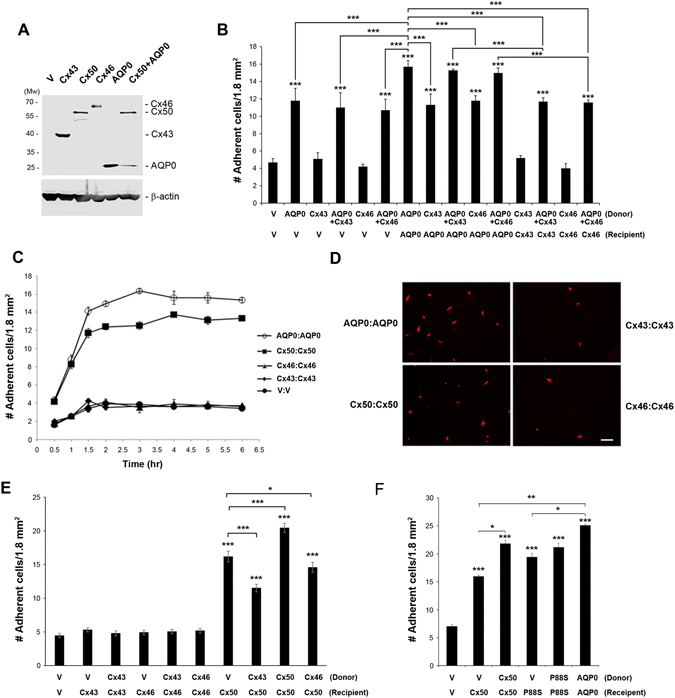
Connexins play essential roles in lens homeostasis and development. Here, we identified a new role for Cx50 that mediates cell-cell adhesion function. Cx50 enhanced the adhesive capability of AQP0. Interestingly, the expression of Cx50 alone promoted cell adhesion at a comparable level to AQP0; however, this cell adhesive function was not observed with other lens connexins, Cx43 and Cx46. Moreover, the adhesive property occurred in both homotypic with Cx50 expressed in both pairing cells and heterotypic with Cx50 in only one pairing cell, and this function appears to be unrelated to its role in forming gap junction channels. Cx50 KO lenses exhibited increased intercellular spaces between lens fiber cells. The second extracellular loop domain (E2) is primarily responsible for this adhesive function. Treatment with a fusion protein containing E2 domain inhibited cell adhesion. Furthermore, disruption of cell adhesion by the E2 domains impaired primary lens cell differentiation. Five critical amino acid residues in the E2 domain primarily are involved in cell adhesive function as well as lens epithelial-fiber differentiation. Together, these results suggest that in addition to forming gap junction channels, Cx50 acts as an adhesive molecule that is critical in maintaining lens fiber integrity and epithelial-fiber differentiation.


Conflict of interest statement

The authors declare that they have no competing interests.

Figures

Figure 1
Figure 1
Cx50 increases cell-cell adhesion and enhances the adhesive capability of AQP0. (A) Crude membrane extracts were prepared from CEF cells infected with high titer RCAS(A) retroviral vehicle (V) or recombinant RCAS(A) retroviruses containing Cx50, AQP0 or co-infected with both Cx50 and AQP0 and were immunoblotted with anti-Flag or β-actin antibody. (B) The diagram illustrates cell adhesion assay via “parachuting” Dil-fluorescence-labeled donor cells onto the confluent recipient cells. (C–E) High titer recombinant retroviruses containing RCAS(A) vehicle (V), Cx50 or AQP0 were used to infect CEF cells. Cell-cell adhesion assay with donor and receipt cells infected with various combinations of recombinant retroviruses containing Cx50 or/and AQP0. After 1.5 hr incubation, fluorescent adherent donor cells were counted and quantified. The data are presented as the mean ± SEM. n = 3. As compared to vehicle controls (V), ***P < 0.001.
Figure 2
Figure 2
Cx50, not the other two lens connexins, Cx43 and Cx46, mediates cell-cell adhesion, and cell adhesion function is independent of Cx50 channel-forming function. Retroviruses containing RCAS(A) vehicle (V), Cx50, Cx46, Cx43 or AQP0 were used to infect CEF cells. (A) Crude membrane extracts of CEF cells expressing various connexins or/and AQP0 were immunoblotted with anti-Flag or β-actin antibody. (B) Cell-cell adhesion assay was conducted with donor or recipient cells expressing various combinations of connexins and AQP0. After 1.5 hr incubation, adherent cells were counted and quantified. (C) Cell adhesion assay was performed in CEF cells homotypic expressing AQP0, Cx50, Cx43, Cx46 or vehicle (V) control after various time periods of incubation. Adherent cells were counted and quantified. (D) Representative fluorescence images of adherent cells are shown in red fluorescence. Bar, 50 µm. (E) Cell adhesion assay was performed in CEF cells expressing lens connexins or vehicles (V) homotypic or heterotypic. (F) High titter recombinant retroviruses containing RCAS(A) vehicle (V), Cx50, Cx50P88S mutant or AQP0 were used to infect CEF cells. Cell adhesion assay was conducted and adherent cells were counted and quantified. The data are presented as the mean ± SEM. n = 3. As compared to vehicle (V) controls or otherwise indicated, *P < 0.05; **P < 0.01; ***P < 0.001.
Figure 3
Figure 3
Second extracellular loop domain (E2) Cx50 mediates the adhesion function of Cx50 and this function is independent of forming functional connexin channels. (A) GST-fusion protein containing E1 and E2 domains were generated and detected by Commassie blue staining. (B) High titter recombinant retroviruses containing RCAS(A) vehicle (V), Cx50 and Cx46 were used to infect CEF cells and cell lysates were prepared. Protein pull down assay was performed using cell lysates and E1-GST, E2-GST or GST attached to glutathione beads. Cell lysates (Lysates) and elutes (E1-GST, E2-GST or GST) from the beads were immunoblotted with anti-FLAG antibody. (C) High titter recombinant retroviruses containing RCAS(A) vehicle (V), Cx50, Cx50P88S mutant or AQP0 were used to infect CEF cells in the absence or presence of GST-fusion protein containing E1 or E2 domain. Cell adhesion assay was conducted and adherent cells were counted and quantified. The data are presented as the mean ± SEM. n = 3. ***P < 0.001.
Figure 4
Figure 4
Cx50 mediates cell adhesion function and Cx50 deficiency in lens fiber cells resulted in increased intercellular spaces. (A) High titter recombinant lentivirus containing vehicle (V) or Cx50 were used to infect HEK293 cells. Cell adhesion assay was conducted and adherent cells were counted and quantified. The data are presented as the mean ± SEM. n = 3. **P < 0.01; ***P < 0.001. (B) H&E staining of paraffin tissue sections of postnatal day 15 mouse lenses of WT or Cx50 KO mice were prepared and representative images were taken from the anterior cortex of the lenses. Bar, 20 µm. n ≥ 3. (C) One month old mouse lenses of WT or Cx50 gene KO mice were processed for thin section TEM. Representative images from the anterior superficial cortex of the lenses show that cortical fiber cells exhibit the intact cell membranes (arrows) in WT, but display enlarged intercellular spaces of various sizes in single KO lenses for Cx50. Several short intact cell membranes (arrows) were also observed among these enlarged spaces. All scale bars, 500 nm. n ≥ 3.
Figure 5
Figure 5
Disruption of cell adhesion by Cx50 E2 domain impairs lens epithelial-fiber cell differentiation. (A) Cx50 E1-GST, E2-GST or GST was used to treat primary lens cell culture every day starting at the 2nd day of cell seeding. Representative images of primary culture cells treated with GST-E1, E2 fusion proteins or GST protein and untreated control were taken on the 10th day of primary culturing. Starting at the 6th day of the treatment, the total lentoid numbers were quantified each day until day 14. (B) Fourteen days after injection, fixed primary lens cells were immunostained with anti-AQP0 antibody and detected by fluorescein-conjugated anti-rabbit IgG. The signals of AQP0 expression areas versus whole-image areas were quantified. The data are presented as the mean ± SEM. *P < 0.05; **P < 0.01; ***P < 0.001. n = 3. Bar, 200 µm.
Figure 6
Figure 6
Identification of critical amino acid residues at E2 domain involved in cell adhesion and lens epithelial-fiber differentiation. (A) GST-fusion proteins containing WT E2, and various single site mutation of E2 domain were generated and detected by Commassie blue staining. (B) High titter recombinant retroviruses containing RCAS(A) vehicle (V) and Cx50, were used to infect CEF cells in the absence or presence of GST-fusion protein containing WT or 8 single site-mutated E2 domain. Cell adhesion assay was conducted and adherent cells were counted and quantified. The data are presented as the mean ± SEM. n = 3. As compared to untreated controls, *P < 0.05; ***P < 0.001. (C) GST fusion proteins containing WT or single site mutated E2 domain or GST was used to treat primary lens cell culture every day starting at the 2nd day of cell seeding. Fourteen days after injection, fixed primary lens cells were immunostained with anti-AQP0 antibody and detected by fluorescein-conjugated anti-rabbit IgG. The signals of AQP0 expression areas versus whole-image areas were quantified. The data are presented as the mean ± SEM. n = 3. As compared to untreated control (C), ***P < 0.001.

Similar articles

Cited by

References

    1. Mese G, Richard G, White TW. Gap junctions: Basic structure and function. J Invest Dermatol. 2007;127:2516–2524. doi: 10.1038/sj.jid.5700770. - DOI - PubMed
    1. Gerido DA, White TW. Connexin disorders of the ear, skin, and lens. Biochim Biiophys Acta. 2004;1662:159–170. doi: 10.1016/j.bbamem.2003.10.017. - DOI - PubMed
    1. Gong X, Cheng C, Xia C. Connexins in lens development and cataractogenesis. J Memb Biol. 2007;218:9–12. doi: 10.1007/s00232-007-9033-0. - DOI - PubMed
    1. Yu XS, Jiang JX. Interaction of major intrinsic protein (aquaporin-0) with fiber connexins in lens development. J Cell Sci. 2004;117:871–880. doi: 10.1242/jcs.00945. - DOI - PubMed
    1. Liu J, et al. Aquaporin 0 enhances gap junction coupling via its cell adhesion function and interaction with connexin 50. J Cell Sci. 2011;124:198–206. doi: 10.1242/jcs.072652. - DOI - PMC - PubMed

Publication types