. 2017 Jul 13;7(1):5303.
doi: 10.1038/s41598-017-05525-4.

A Simple Add-and-Display Method for Immobilisation of Cancer Drug on His-tagged Virus-like Nanoparticles for Controlled Drug Delivery

Affiliations

A Simple Add-and-Display Method for Immobilisation of Cancer Drug on His-tagged Virus-like Nanoparticles for Controlled Drug Delivery

Roya Biabanikhankahdani et al. Sci Rep. .

Abstract

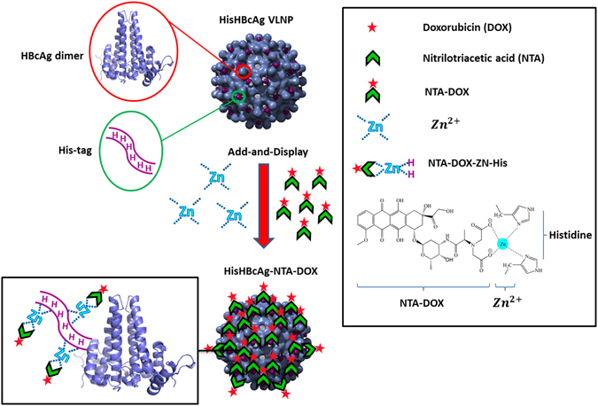
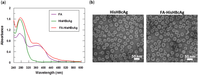
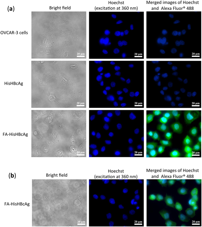
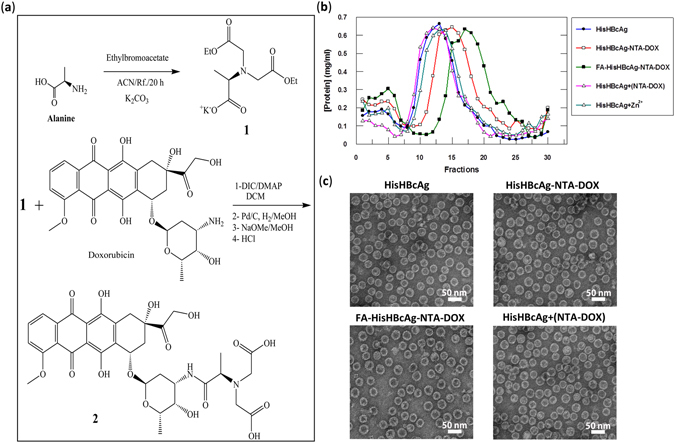
pH-responsive virus-like nanoparticles (VLNPs) hold promising potential as drug delivery systems for cancer therapy. In the present study, hepatitis B virus (HBV) VLNPs harbouring His-tags were used to display doxorubicin (DOX) via nitrilotriacetic acid (NTA) conjugation. The His-tags served as pH-responsive nanojoints which released DOX from VLNPs in a controlled manner. The His-tagged VLNPs conjugated non-covalently with NTA-DOX, and cross-linked with folic acid (FA) were able to specifically target and deliver the DOX into ovarian cancer cells via folate receptor (FR)-mediated endocytosis. The cytotoxicity and cellular uptake results revealed that the His-tagged VLNPs significantly increased the accumulation of DOX in the ovarian cancer cells and enhanced the uptake of DOX, which improved anti-tumour effects. This study demonstrated that NTA-DOX can be easily displayed on His-tagged VLNPs by a simple Add-and-Display step with high coupling efficiency and the drug was only released at low pH in a controlled manner. This approach facilitates specific attachment of any drug molecule on His-tagged VLNPs at the very mild conditions without changing the biological structure and native conformation of the VLNPs.


Conflict of interest statement

The authors declare that they have no competing interests.

Figures

Figure 1
Figure 1
Schematic representation of the Add-and-Display method for immobilisation of doxorubicin non-covalently on His-tagged VLNPs. The HisHBcAg VLNP is made up of many copies of HBcAg dimers (blue). The His-tag fused at the N-terminal end of the HBcAg monomer forms trimeric spikes (magenta) and are exposed on the surface of the HisHBcAg VLNP. NTA-DOX is synthesised from nitrilotriacetic acid and doxorubicin hydrochloride. In the presence of Zn2+, the NTA-DOX interacts with histidine residues and is displayed on the surface of the VLNP.
Figure 2
Figure 2
Conjugation of folic acid to His-tagged VLNPs. (a) Spectra of folic acid (FA), HisHBcAg nanoparticles (HisHBcAg), and FA-conjugated HisHBcAg nanoparticles (FA-HisHBcAg). (b) Electron micrographs of HisHBcAg nanoparticles. The samples (as labelled on top of the micrographs) were stained with uranyl acetate and viewed under a TEM. All the samples assembled into spherical structures. White bars indicate 50 nm.
Figure 3
Figure 3
Internalisation of His-tagged VLNPs into OVCAR-3 cells. The HisHBcAg nanoparticles (HisHBcAg; 25 μg) and folic acid (FA)-conjugated HisHBcAg nanoparticles (FA-HisHBcAg; 25 μg) were incubated with OVCAR-3 cells for 16 h at 37 °C. The internalised HisHBcAg nanoparticles were detected by the rabbit anti-HBcAg serum, followed by anti-rabbit antibody conjugated to Alexa Fluor® 488 (a), and the mouse anti-His antibody, followed by anti-mouse antibody conjugated to Alexa Fluor® 488 (b). Cell nuclei were stained with Hoechst 33342. Non-transfected cancer cells served as a negative control.
Figure 4
Figure 4
Synthesis and immobilisation of NTA-DOX on His-tagged VLNPs. (a) Synthesis of NTA-DOX. The amine group of alanine (Ala) was fully protected with ethylbromoacetate using K2CO3 under reflux condition to obtain diester-Ala (compound 1). Then, the free carboxylic acid of diester-Ala was activated by N,N′-diisopropylcarbodiimide (DIC) and 4-Dimethylaminopyridine (DMAP). The activated diester-Ala was reacted with doxorubicin (DOX) to produce diester-Ala-DOX, which was then converted to dicarboxylic-Ala-DOX (NTA-DOX; compound 2) by the hydrolysis method using NaOMe in methanol. (b) The NTA-DOX was incubated with the HisHBcAg nanoparticles (HisHBcAg) and folic acid (FA)-conjugated HisHBcAg nanoparticles (FA-HisHBcAg) in the presence of Zn2+. The nanoparticles conjugated with NTA-DOX were purified by sucrose density gradient ultracentrifugation. The protein amount in each fraction (400 μL) was determined using the Bradford assay. The HisHBcAg nanoparticles (HisHBcAg), HisHBcAg nanoparticles incubated with NTA-DOX in the absence of Zn2+ [HisHBcAg + (NTA-DOX)] and HisHBcAg nanoparticles incubated with Zn2+ (HisHBcAg + Zn2+) served as negative controls. (c) Electron micrographs of different HisHBcAg VLNPs formed by HisHBcAg, HisHBcAg-NTA-DOX, FA-HisHBcAg-NTA-DOX, and HisHBcAg + (NTA-DOX). White bars indicate 50 nm.
Figure 5
Figure 5
Identification of doxorubicin immobilised on His-tagged VLNPs. (a and b) Doxorubicin (DOX) was detected by measuring absorbance at 495 nm of the fractions obtained from sucrose gradients. (a) DOX, NTA-DOX, HisHBcAg nanoparticles incubated with NTA-DOX in the absence of Zn2+ [HisHBcAg + (NTA-DOX)] stayed on top of the sucrose gradients after centrifugation. HisHBcAg nanoparticles (HisHBcAg) were not detected at A495. HisHBcAg nanoparticles incubated with Zn2+ (HisHBcAg + Zn2+) showed a negligible absorbance at 495 nm. (b) HisHBcAg nanoparticles conjugated non-covalently with NTA-DOX (HisHBcAg-NTA-DOX) and HisHBcAg nanoparticles conjugated covalently with folic acid (FA) and non-covalently with NTA-DOX (FA-HisHBcAg-NTA-DOX) migrated into the sucrose gradient, and the immobilised DOX was detected with A495. (c and d) Native agarose gel electrophoresis of the HisHBcAg nanoparticles conjugated non-covalently with DOX. The same gel was visualised under (c) ultraviolet (UV) illumination, and (d) stained with Coomassie Brilliant Blue (CBB).
Figure 6
Figure 6
Doxorubicin release profile of the His-tagged VLNPs at different pH. The release profiles of free doxorubicin (DOX), HisHBcAg nanoparticles conjugated non-covalently with NTA-DOX (HisHBcAg-NTA-DOX) and HisHBcAg nanoparticles conjugated covalently with folic acid (FA) and non-covalently with NTA-DOX (FA-HisHBcAg-NTA-DOX) at pH 5.4 and 7.4. More than 80% of the free DOX was released after 5 h at pH 5.4 and pH 7.4, whereas approximately 80% of the conjugated DOX on HisHBcAg nanoparticles was released after 16 h at pH 5.4. Data are expressed as mean ± standard deviation (n = 3).
Figure 7
Figure 7
Localisation of His-tagged VLNPs in cancer and normal cells by live cell imaging microscopy. (a) Ovarian cancer OVCAR-3 and (b) normal 3T3 cells were incubated with free doxorubicin (DOX), HisHBcAg nanoparticles conjugated non-covalently with NTA-DOX (HisHBcAg-NTA-DOX) and HisHBcAg nanoparticles conjugated covalently with folic acid (FA) and non-covalently with NTA-DOX (FA-HisHBcAg-NTA-DOX) at equivalent DOX concentration (5 μg/mL) for 1 h at 37 °C. The untreated cells served as negative controls. Cell nuclei were stained with Hoechst 33342, and DOX was excited at 480 nm and emitted at 535 nm. The samples are labelled on the left. Scale bars indicate 20 μm.
Figure 8
Figure 8
Cytotoxicity analysis of various doxorubicin formulations on ovarian cancer and normal cells. Viability of ovarian cancer OVCAR-3 (a), and normal 3T3 (b) cells. The HisHBcAg nanoparticles conjugated covalently with folic acid (FA) and non-covalently with NTA-DOX (FA-HisHBcAg-NTA-DOX) were more efficient in inhibiting the growth of OVCAR-3 cells compared to that of other formulations. On the other hand, this formulation (FA-HisHBcAg-NTA-DOX) was less toxic to normal 3T3 cells, resulted in a conferred protection of the normal cells from DOX. The HisHBcAg nanoparticles (HisHBcAg) were not toxic to both normal and cancer cells as shown in the small graphs on the right. Data represent mean ± SD of triplicate determinations.

Similar articles

Cited by

References

    1. Liu J, et al. pH-sensitive nano-systems for drug delivery in cancer therapy. Biotechnol. Adv. 2014;32:693–710. doi: 10.1016/j.biotechadv.2013.11.009. - DOI - PubMed
    1. Liu Y, Wang W, Yang J, Zhou C, Sun J. pH-sensitive polymeric micelles triggered drug release for extracellular and intracellular drug targeting delivery. Asian J. Pharm. Sci. 2013;8:159–167. doi: 10.1016/j.ajps.2013.07.021. - DOI
    1. Karimi M, et al. pH‐Sensitive stimulus‐responsive nanocarriers for targeted delivery of therapeutic agents. Wiley Interdiscip. Rev. Nanomed. Nanobiotechnol. 2016;8:696–716. doi: 10.1002/wnan.1389. - DOI - PMC - PubMed
    1. Karimi M, et al. Smart micro/nanoparticles in stimulus-responsive drug/gene delivery systems. Chem. Soc. Rev. 2016;45:1457–1501. doi: 10.1039/C5CS00798D. - DOI - PMC - PubMed
    1. Wang S, Huang P, Chen X. Stimuli-responsive programmed specific targeting in nanomedicine. ACS Nano. 2016;10:2991–2994. doi: 10.1021/acsnano.6b00870. - DOI - PMC - PubMed

Publication types

LinkOut - more resources