. 2017 Jul 13;7(1):5296.
doi: 10.1038/s41598-017-05680-8.

DGKδ triggers endoplasmic reticulum release of IFT88-containing vesicles destined for the assembly of primary cilia

Affiliations

DGKδ triggers endoplasmic reticulum release of IFT88-containing vesicles destined for the assembly of primary cilia

Jie Ding et al. Sci Rep. .

Abstract

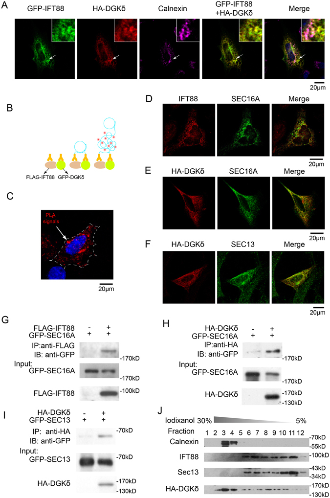
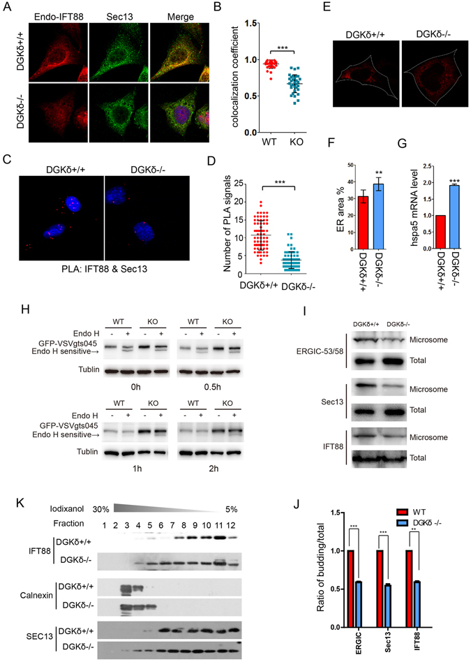
The morphogenic factor Sonic hedgehog (Shh) signals through the primary cilium, which relies on intraflagellar transport to maintain its structural integrity and function. However, the process by which protein and lipid cargos are delivered to the primary cilium from their sites of synthesis still remains poorly characterized. Here, we report that diacylglycerol kinase δ (DGKδ), a residential lipid kinase in the endoplasmic reticulum, triggers the release of IFT88-containing vesicles from the ER exit sites (ERES), thereby setting forth their movement to the primary cilium. Encoded by the gene whose mutations originally implicated the primary cilium as the venue of Shh signaling, IFT88 is known to be part of the complex B that drives the anterograde transport within cilia. We show that IFT88 interacts with DGKδ, and is associated with COPII-coated vesicles at the ERES. Using a combination of RNAi silencing and gene knockout strategies, we further show that DGKδ is required for supporting Shh signaling both in vitro and in vivo, demonstrating the physiological significance of this regulation.


Conflict of interest statement

The authors declare that they have no competing interests.

Figures

Figure 1
Figure 1
IFT88 is a component of ER-derived COPII coated vesicles. (A) Representative immunofluorescence staining images of endogenous IFT88 and SEC13 in the perinuclear region of NIH3T3 cells. The Manders Overlap Coefficient (MOC) between IFT88 and SEC13 is 0.72 ± 0.093. (B) Representative immunofluorescence confocal images showing colocalization of exogenously expressed GFP-IFT88 with endogenous Sec31A. MOC, 0.61 ± 0.03. (C and D) Representative confocal images showing colocalization of endogenous IFT88 with exogenously expressed ER marker DsRed-ER or exogenously expressed GFP-IFT88 with endo-Calnexin. Arrows mark the positions of the insets. MOC, 0.55 ± 0.11 in (C) and 0.68 ± 0.085 in (D), respectively. (E) Western analysis of IFT88 and SEC13 present in four differential centrifugation fractions in NIH3T3 cells. (F and G) Co-immunoprecipitation of exogenously expressed FLAG-IFT88 and GFP-Sec13 (F) or FLAG-IFT88 and CFP-Sec31A (G) in 293T cells. The cell lysates were precipitated with anti-FLAG M2 affinity gel and probed with anti-GFP or anti-Sec31A antibody for Western blot. The statistics analysis was computed based on data from at least 3 independent experiments.
Figure 2
Figure 2
Identification of DGKδ as a specific IFT88 interacting protein. (A) Silver staining of proteins immunoprecipitated with anti-IFT88 or normal IgG control from NIH3T3 cell lysates and resolved by PAGE. 3 unique bands enriched in the anti-IFT88 lane were excised and identified by mass spectrometry. (B) Results from mass spectrometry analysis showing the identity of 3 bands to be: DGKδ, IFT81 and Klra19, respectively. (C) IP-Western analysis of exogenously expressed FLAG-tagged IFT88 and HA-tagged DGK isoforms in 293T cells. Top panel, anti-FLAG blotting after anti-HA immunoprecipitation. Total controls are shown in the bottom two panels. (D) Schematic representation of IFT88 domain structure and deletion constructs. (E) IP-Western analysis of the interaction between DGKδ and various IFT88 constructs. Total controls are shown in the bottom two panels. (F) Schematic representation of DGKδ domain structure and deletion constructs. (G) IP-Western analysis of the interaction between IFT88 and various DGKδ constructs.
Figure 3
Figure 3
IFT88 is associated with COPII vesicles at the ER exit sites. (A) Representative immunofluorescence images of exogenously expressed GFP-IFT88, HA-DGKδ as well as endogenous calnexin in NIH3T3 cells. Arrows mark the positions of insets. MOC between IFT88 and DGKδ: 0.89 ± 0.065. (B) Schematic diagram and (C) a representative image of results from the proximity ligation assay showing that exogenously expressed FLAG-IFT88 and pEGFP-DGKδ are colocalized in the peri-nuclear region in NIH3T3 cells (the red signals adjacent came from another transfected cell). (D–F) Representative immunofluorescence images showing double staining of GFP-SEC16A and IFT88 (D), HA-DGKδ and GFP-SEC16A (E), as well as HA-DGKδ and SEC13 (F), respectively, in NIH3T3 cells. MOC: 0.68 ± 0.053 in (D), 0.81 ± 0.042 in (E), 0.63 ± 0.066 in (F). (G) IP-Western analysis of the interaction between exogenously expressed FLAG-IFT88 and GFP-SEC16A, (H) HA-DGKδ and GFP-SEC16A, as well as (I) HA-DGKδ and GFP-SEC13 in HEK293T cells. (J) Iodixanol density gradient sedimentation experiments in HEK293T cells, in which samples from 12 fractions were analyzed by Western blot for calnexin, IFT88, SEC13, and exogenously expressed HA-DGKδ. These experiments were reproduced in at least 3 independent experiments.
Figure 4
Figure 4
DGKδ triggers ER release of IFT88-containing COPII vesicles. (A) Representative immunostaining images of endogenous IFT88 and SEC13 in DGKδ−/− and their matching control (DGKδ+/+) MEFs. (B) Calculation of colocalization co-efficient in (A). (C) Proximity ligation assay for and (D) quantification of the interaction between IFT88 and SEC13 in DGKδ −/− MEFs and the control (DGKδ+/+) MEFs. Data are means plus standard deviation and the statistical significance was calculated with the Student T test, n > 40. (E) Representative images and (F) quantification of ER areas in DGKδ+/+ and DGKδ−/− MEFs. The cells were marked by exogenously expressed DsRed-ER, and the white dotted lines mark the contour of the cell. Data are means plus standard deviation and the statistical significance was calculated with the Student T test, n > 20. (G) Real-time qPCR analysis of Hspa5 mRNA in DGKδ+/+ and DGKδ−/− MEFs. (H) Westernblot analyses Endo H sensitive of VSVgts045 in DGKδ+/+ and DGKδ−/− MEFs. The cells were routinely maintained at the permissive 37 °C, shifted to the nonpermissive 40 °C for 24 hours to let GFP-VSVgts045 accumulate in the ER after transfection with a plasmid vector expressing GFP-VSVgts045, then added cycloheximide to inhibit protein synthesis and shifted back to permissive 32 °C for various time periods to monitor VSVgts045 trafficking after adding cycloheximide to inhibit protein synthesis. At the end of each time point, the cells were harvested for Endo H digestion and analyzed by westernblot. (I) Western blot results showing the distribution of ERGIC53, IFT88 and Sec13 in COP II-coated vesicles which budding in vitro. And the distribution of the three proteins and β-actin were taken as the control. (J) Bar graph depicts the ratio of relative intensity of ERGIC53, Sec13 and IFT88 in budding and total portions in DGKδ+/+ and DGKδ−/− MEFs in (I). (K) Iodixanol density gradient sedimentation of calnexin, IFT88, SEC13, and exogenously expressed HA-DGKδ in DGKδ+/+ and DGKδ−/− MEFs. **P < 0.01, ***P < 0.001.
Figure 5
Figure 5
DGKδ is required for maintaining cilium length and IFT transport. (A) Immunofluorescence staining of IFT88(green) and acetylated tubulin (red) in DGKδ+/+ and DGKδ−/− MEFs. The cells were allowed to reach confluency, then serum starved for 24 hours before being analyzed. (B) Quantification of cilium length and (C) intensity of IFT88 staining in (A) are presented as means ± SD, N > 20. (D) Quantification of cilium length in DGKδ−/− MEFs transfected with pEGFP, pEGFP-DGKδ and pEGFP-DGKδG337D. The cells were serum-starved post-transfection for 24 hours to allow for cilium formation. N > 20. (E–F) FRAP measurement of IFT88-GFP import into the primary cilium in DGKδ+/+ and DGKδ−/− MEFs. The cells were treated as in (D), and images of fluorescence recovery were captured every 30 s after photobleaching in the entire cilium. (G) Immunofluorescence measurement and (H) quantification of exogenously expressed GFP-Peripherin/rds and GFP-SSTR3 in primary cilia of DGKδ+/+ and DGKδ−/− MEFs. The cells were stained with anti-Ac-tubulin to mark the axoneme of cilia. Data are means plus SD, and statistical analysis was carried out with Student’s T test. **P < 0.01, ***P < 0.001.
Figure 6
Figure 6
DGKδ is required for supporting Shh signaling functions. (A) Real-time qPCR analyses of Gli1 mRNA in NIH3T3 transfected with various siRNAs as indicated. ShhN-CM (1:10) was given for 12 hours. (B) Real-time PCR analyses of siDGKδ-1 and siDGKδ-2 knockdown efficiency in NIH3T3. (C,D) 8xGBS-luc reporter assays for the ability of DGK inhibitor R59022 (C) and R59949 (D) to inhibit Shh signaling induced by ShhN-CM (1:10) or SAG. (E) Western analysis of Gli1 protein level induced by ShhN or SAG in the absence or presence of R59022 (10 μM) or R59949 (7.5 μM). (F) Quantification of Gli1 intensity in (E). (G) Representative confocal images of Smo-GFP in cilia of Smo−/− MEFs, in which Smo-GFP was re-introduced by stable transfection. The treatment with ShhN-CM and R59022 or R59949 was given for 4 hours. (H) Quantification of Smo-GFP intensity in (G), N > 20. (I) Representative images of immunofluorescence staining and (J) quantification of Gli3 in cilia of NIH3T3 cells. The treatment was given as in (G). (K) Quantification of EdU incorporation by freshly isolated cerebellar GCP cells. The cells were transfected with non-targeting siIFT88, siDGKδ-1, siDGKδ-2, or a non-silencing control (NS), and treated with or without ShhN as indicated.
Figure 7
Figure 7
A model for IFT88 mediated secretory pathway from ER to primary cilia. IFT88 is found at the ER exit sites, where it is incorporated into the ER-derived COP II-coated vesicles. The IFT88 containing COP II vesicles travel directly to cilia bypassing the Golgi apparatus. The newly identified IFT88-binding protein DGKδ is colocalized with IFT88 at the ER exit sites and is required for triggering the release of IFT88 vesicles from the ER.

References

    1. Jiang J, Hui CC. Hedgehog signaling in development and cancer. Developmental cell. 2008;15:801–812. doi: 10.1016/j.devcel.2008.11.010. - DOI - PMC - PubMed
    1. Cohen MM., Jr. Hedgehog signaling update. American journal of medical genetics. Part A. 2010;152a:1875–1914. doi: 10.1002/ajmg.a.32909. - DOI - PubMed
    1. Huangfu D, et al. Hedgehog signalling in the mouse requires intraflagellar transport proteins. Nature. 2003;426:83–87. doi: 10.1038/nature02061. - DOI - PubMed
    1. Corbit KC, et al. Vertebrate Smoothened functions at the primary cilium. Nature. 2005;437:1018–1021. doi: 10.1038/nature04117. - DOI - PubMed
    1. Goetz SC, Anderson KV. The primary cilium: a signalling centre during vertebrate development. Nature reviews. Genetics. 2010;11:331–344. doi: 10.1038/nrg2774. - DOI - PMC - PubMed

Publication types

Substances

LinkOut - more resources