. 2015 Mar 1;6(3):1770-1779.
doi: 10.1039/c4sc02340d. Epub 2014 Nov 26.

Protein ubiquitination and formation of polyubiquitin chains without ATP, E1 and E2 enzymes

Affiliations

Protein ubiquitination and formation of polyubiquitin chains without ATP, E1 and E2 enzymes

Sungjin Park et al. Chem Sci. .

Abstract

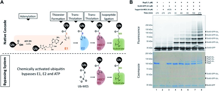
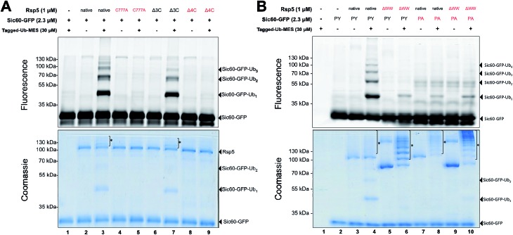
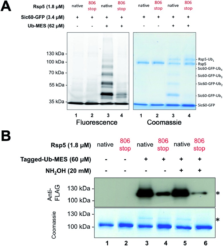
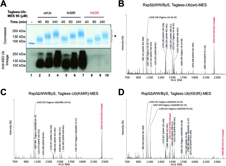
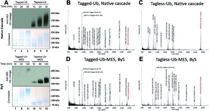
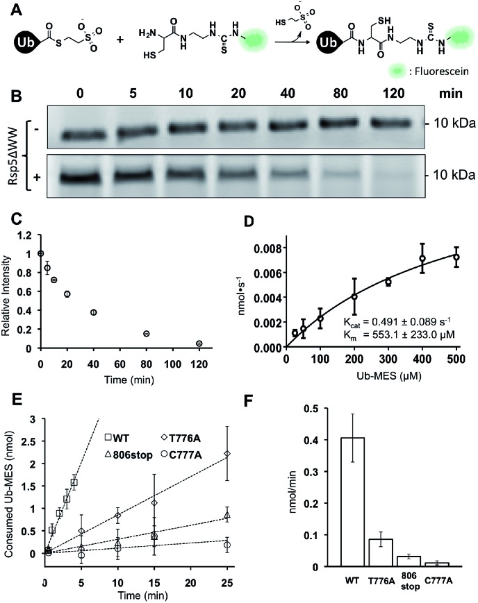
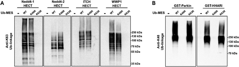
Studying protein ubiquitination is difficult due to the complexity of the E1-E2-E3 ubiquitination cascade. Here we report the discovery that C-terminal ubiquitin thioesters can undergo direct transthiolation with the catalytic cysteine of the model HECT E3 ubiquitin ligase Rsp5 to form a catalytically active Rsp5∼ubiquitin thioester (Rsp5∼Ub). The resulting Rsp5∼Ub undergoes efficient autoubiquitination, ubiquitinates protein substrates, and synthesizes polyubiquitin chains with native Ub isopeptide linkage specificity. Since the developed chemical system bypasses the need for ATP, E1 and E2 enzymes while maintaining the native HECT E3 mechanism, we named it "Bypassing System" (ByS). Importantly, ByS provides direct evidence that E2 enzymes are dispensable for K63 specific isopeptide bond formation between ubiquitin molecules by Rsp5 in vitro. Additionally, six other E3 enzymes including Nedd4-1, Nedd4-2, Itch, and Wwp1 HECT ligases, along with Parkin and HHARI RBR ligases processed Ub thioesters under ByS reaction conditions. These findings provide general mechanistic insights on protein ubiquitination, and offer new strategies for assay development to discover pharmacological modulators of E3 enzymes.


Figures

Fig. 1
Fig. 1. Bypassing System (ByS). (A) C-terminal ubiquitin thioester Ub–MES can form a catalytically active HECT E3∼Ub thioester adduct and conjugate ubiquitin to protein substrates, bypassing ATP, E1, and E2. (B) A time course of Sic60-GFP ubiquitination by Rsp5 and Tagged-Ub–MES. Reaction mixtures were incubated at room temperature for indicated times, quenched with Laemmli buffer, resolved by SDS-PAGE and imaged with in-gel fluorescence scanning and coomassie staining.
Fig. 2
Fig. 2. Ubiquitination via Rsp5/ByS depends on the catalytic cysteine of Rsp5 and enzyme/substrate recognition. (A) Rsp5/ByS depends on the presence of the Rsp5 catalytic cysteine. Ubiquitination of Sic60-GFP by Rsp5 mutants via ByS was analyzed by in-gel fluorescence scanning. Rsp5 variants without the catalytic cysteine (Cys777) are colored red. Autoubiquitinated Rsp5 is marked with *. Δ3C:Rsp5 lacks three non-catalytic cysteines. Δ4C:Rsp5 lacks all four cysteines. (B) Ubiquitination of Sic60-GFP via Rsp5/ByS depends on enzyme–substrate recognition. Ubiquitination of Sic60-GFP or Sic60PA-GFP in the presence of Rsp5 or Rsp5ΔWW was analyzed by in-gel fluorescence scanning. Rsp5ΔWW and Sic60PA-GFP are colored in red. Autoubiquitinated Rsp5 and Rsp5ΔWW are marked with *. ΔWW:Rsp5 lacks its WW domains, which recognize PY motifs on the substrate. PY: Sic60-GFP has its PY motif, while Sic60PA-GFP has a PY → PA mutation.
Fig. 3
Fig. 3. Ubiquitination via Rsp5/ByS recapitulates the instrinsic mechanism of Rsp5. (A) Ubiquitination of Sic60-GFP via Rsp5/ByS depends on the last four C-terminal amino acids of Rsp5. The ubiquitination of Sic60-GFP via Rsp5806stop/ByS is analyzed by in-gel fluorescence scanning after 4 hours of reaction time. (B) Rsp5806stop forms an inactive thioester adduct with Tagged-Ub-MES (Rsp5806stop∼Tagged-Ub). The formation of Rsp5806stop∼Tagged-Ub in the Rsp5806stop/ByS reaction was confirmed by Western-blotting with anti-FLAG antibody and coomassie staining. Reaction mixtures were incubated for 45 minutes at room temperature and quenched by either non-reducing Laemmli buffer or reducing Laemmli buffer containing NH2OH (20 mM, final concentration). The band corresponding to Rsp5806stop∼Tagged-Ub adduct is marked with *.
Fig. 4
Fig. 4. Rsp5/ByS forms K63-linked polyubiquitin chains. (A) K63-linked polyubiquitin chains formed by Rsp5ΔWW and Tagless-Ub–MES were visualized by western-blotting with K63-ubiquitin linkage specific antibodies. A mixture of Rsp5ΔWW (1.8 μM) and each Ub–MES mutant was incubated for indicated times and analyzed by coomassie staining and western-blotting. MALDI-TOF analysis was performed for the higher MW bands produced in the reaction with Rsp5ΔWW and (B) Tagless-Ub(wt)–MES (lane 4), (C) Tagless-Ub(K48R)–MES (lane 7), and (D) Tagless-Ub(K63R)–MES (lane 10). The gel region of 100–250 kDa from lane 4, 7, and 10 was excised for MALDI-TOF analysis. Any peak corresponding to the calculated polyubiquitin linkage signal (Table S2–S4†) is marked in red. Autoubiquitinated Rsp5ΔWW is marked with *.
Fig. 5
Fig. 5. N-terminal tag on ubiquitin (Tagged-Ub) interferes with the formation of K63-polyubiquitin chains by Rsp5ΔWW under native and ByS reaction conditions. (A) The presence of K63-linkages in polyubiquitin chains formed by Rsp5 with Tagged- and Tagless-ubiquitin under both native cascade and ByS conditions was detected with western-blotting using anti-K63-linkage antibody. Native ubiquitination reactions contained UBE1 (0.09 μM), UbcH5a (1.0 μM), hydrolyzed Tagged- or Tagless-ubiquitin (90 μM), Rsp5ΔWW (2.0 μM) and ATP (4 mM). For ByS reactions, Rsp5ΔWW (2.0 μM) was treated with Tagless-Ub–MES (50 μM) or Tagged-Ub–MES (40 μM) and incubated at room temperature for the indicated times. MALDI-TOF analysis was performed for polyubiquitin chains formed by Rsp5ΔWW with (B) Tagged-Ub via native cascade (lane 3), (C) Tagless-Ub via native cascade (lane 6), (D) Tagged-Ub–MES via ByS (lane 9) and (E) Tagless-Ub–MES via ByS (lane 12). The gel region from 100–250 kDa in lanes 3, 6, 9 and 12 from a coomassie-stained gel was excised, digested by trypsin and analyzed by MALDI-TOF. Any peak corresponding to calculated polyubiquitin linkage signals (ESI Tables S2 and S5†) is marked in red.
Fig. 6
Fig. 6. Kinetic characterization of Rsp5/ByS. (A) Native chemical ligation between Tagless-Ub–MES and FCys. (B) Reaction mixtures were quenched at different time points and the remaining Tagless-Ub–MES was labeled with FCys. Tagless-Ub–MES (250 μM) was incubated for indicated times without Rsp5ΔWW (upper panel) or with Rsp5ΔWW (lower panel, 1.5 μM). (C) Consumption of Tagless-Ub–MES (250 μM) by Rsp5ΔWW (1.5 μM). (D) The initial reaction rates at different Ub–MES concentrations were plotted, and k cat and K m values estimated. (E) Tagless-Ub–MES (250 μM) was incubated with the indicated mutant of Rsp5ΔWW (5 μM) and quenched with FCys at five time points starting at 30 seconds. After linear fitting, the y-intercept of each line was adjusted to equal zero. (F) Comparison of gross consumption rates of Tagless-Ub–MES by Rsp5ΔWW mutants.
Fig. 7
Fig. 7. Activity of HECT E3 ligases and RBR E3 ligases under ByS reaction conditions. (A) The HECT domains of Nedd4-1, Nedd4-2, Itch and Wwp1 (2 μM) were incubated with either Ub(wt)–MES, Ub(K48R)–MES or Ub(K48R)–MES (200 μM) for 90 minutes at room temperature. The reaction mixtures were then analyzed using K63-ubiquitin linkage specific antibody. (B) GST-tagged RBR domains of Parkin (rat) and HHARI (human) (2 μM) were treated with Ub–MES as in (A) and were analyzed using K48-ubiquitin linkage antibody. HHARI and Parkin constructs lack auto-inhibitory domains.

Similar articles

Cited by

References

    1. Varshavsky A. Annu. Rev. Biochem. 2012;81:167. - PubMed
    1. Hershko A., Ciechanover A. Annu. Rev. Biochem. 1998;67:425. - PubMed
    1. Pickart C. M. Annu. Rev. Biochem. 2001;70:503. - PubMed
    1. Deshaies R. J., Joazeiro C. A. Annu. Rev. Biochem. 2009;78:399. - PubMed
    1. Lydeard J. R., Schulman B. A., Harper J. W. EMBO Rep. 2013;14(12):1050–1061. - PMC - PubMed