

doi: 10.1016/j.eats.2017.01.013.
eCollection 2017 Jun.
Basic Hip Arthroscopy: Diagnostic Hip Arthroscopy
Affiliations
- PMID: 28706820
- PMCID: PMC5495576
- DOI: 10.1016/j.eats.2017.01.013
Item in Clipboard
Basic Hip Arthroscopy: Diagnostic Hip Arthroscopy
Arthrosc Tech.
.
Erratum in
-
Erratum.Arthrosc Tech. 2024 Mar 19;13(4):102992. doi: 10.1016/j.eats.2024.102992. eCollection 2024 Apr. Arthrosc Tech. 2024. PMID: 38690344 Free PMC article.
Abstract
Hip arthroscopy is increasing in popularity for the diagnosis and management of hip preservation. The basics of hip arthroscopy positioning, fluoroscopic assessment, and portal establishment are reviewed in the first 2 parts of this series. This article is the third installment in which we describe a systematic approach to performing a diagnostic hip arthroscopy. A mastery of diagnostic arthroscopy is necessary for surgeons treating hip disorders.
Figures
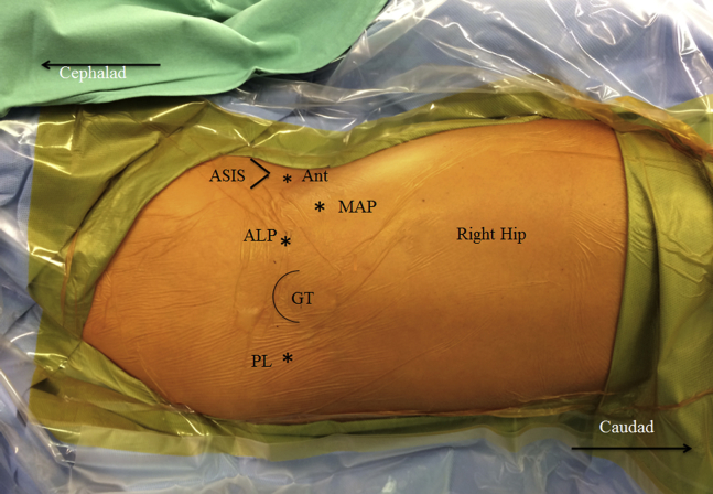
External view depicting anatomic location of bony prominences and arthroscopic portals of a right hip in supine position. The 2 portals used in this surgical technique are the anterolateral portal (AL) and the modified anterior portal (MAP). In 3-portal techniques, the anterior portal (Ant) and/or the posterolateral portal (PL) have traditionally been used. Also depicted here are the bony landmarks used to identify portal location: the anterior superior iliac spine (ASIS) and the greater trochanter (GT).
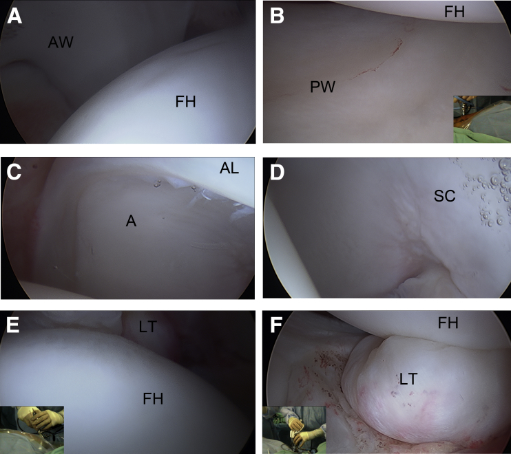
Central compartment viewed from the anterolateral portal of the right hip in the supine position. (A) Visualization of the anterior acetabular wall (AW) with the anterior superior femoral head (FH). (B) Posterior acetabular wall (PW) with the femoral head (FH). (C) 12-o'clock position revealed the acetabulum (A) and anterior labrum (AL). (D) Acetabular stellate crease (SC). (E) View of the fovea, which may identify tears of the ligamentum teres (LT) or incarcerated loose bodies. (F) Ligamentum teres (LT) and the femoral head (FH).
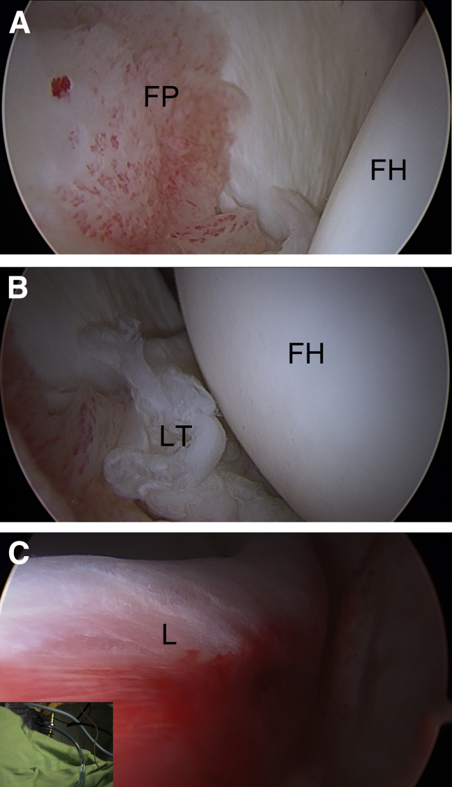
Examples of central compartment pathology of a right hip viewed from modified anterior portal in the supine position. (A) Fat pad inflammation within the cotyloid space. (B) Ligamentum teres (LT) tear. (C) Labral ecchymosis from impingement of the chondrolabral junction. (FH, femoral head; FP, fat pad inflammation; L, labral ecchymosis.)
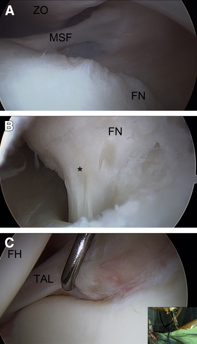
Peripheral compartment of a right hip viewed from the modified anterior portal in the supine position. (A) View of the medial synovial fold, which is an important reference for the medial limit for osteochondroplasty in femoroacetabular impingement. (B) The medial retinacular vessels (*) along the femoral neck (FN). (C) The anterior labral transition to the transverse acetabular ligament (TAL) is revealed by the probe. The picture inlay shows the hand position of the arthroscopy in the modified anterior portal. (FH, femoral head; MSF, medial synovial fold; ZO, zona orbicularis.)
References
-
- Montgomery S.R., Ngo S.S., Hobson T. Trends and demographics in hip arthroscopy in the United States. Arthroscopy. 2013;29:661–665. - PubMed
-
- Bozic K.J., Chan V., Valone F.H., 3rd, Feeley B.T., Vail T.P. Trends in hip arthroscopy utilization in the United States. J Arthroplasty. 2013;28(suppl):140–143. - PubMed
-
- Robertson W.J., Kelly B.T. The safe zone for hip arthroscopy: A cadaveric assessment of central, peripheral, and lateral compartment portal placement. Arthroscopy. 2008;24:1019–1026. - PubMed
LinkOut - more resources
Full Text Sources
Other Literature Sources
