Review
. 2017 Jun;5(12):256.
doi: 10.21037/atm.2017.04.32.

Abdominal aortic aneurysm: pictorial review of common appearances and complications

Affiliations
Review

Abdominal aortic aneurysm: pictorial review of common appearances and complications

Yogesh Kumar et al. Ann Transl Med. 2017 Jun.

Abstract

Abdominal aortic aneurysms (AAAs) are defined as focal dilatations of the abdominal aorta that are 50% greater than the proximal normal segment or when it is more than 3 cm in maximum diameter. The early diagnosis and treatment is very important to prevent catastrophic complications. Due to its ability to assess the peri-aortic soft tissue and the exact extension of aneurysm, as well as its excellent vascular opacification and multiplanar reconstruction capabilities, computed tomography angiography (CTA) has become an integral part of the evaluation of AAA and has virtually replaced conventional angiography for the evaluation of AAA. Knowledge of the characteristic imaging features of AAA is essential for the prompt diagnosis of life-threatening complications. In this pictorial essay, we will discuss the CTA findings in AAA and its complications including rupture, infection, aorto-enteric fistula and aorto-caval fistula.

Keywords: Aortic aneurysm; CT; aorto-caval fistula; aorto-enteric; complications.


Conflict of interest statement

Conflicts of Interest: The authors have no conflicts of interest to declare.

Figures

Figure 1
Figure 1
Plain film of a lumbar spine in a patient with lower back pain (A) demonstrates a large calcified aortic aneurysm (arrow). Follow up CTA and 3D images (B,C,D) demonstrate a large partially thrombosed infrarenal aortic aneurysm (arrow). CTA, computed tomography angiography.
Figure 2
Figure 2
Non-contrast axial (A,B) and coronal (C) CT images of the abdomen demonstrate descending thoracic and abdominal aortic aneurysms with rupture: note the large retroperitoneal hematoma (larger arrow), and periaortic stranding (double arrows). The patient also has a high attenuation crescent sign (smaller arrow)—a secondary sign of rupture.
Figure 3
Figure 3
Axial CTA image demonstrates the presence of high attenuation contrast (larger arrow) in the retroperitoneal hematoma (smaller arrow) suggestive of active bleeding in a ruptured aortic aneurysm. CTA, computed tomography angiography.
Figure 4
Figure 4
Axial and coronal CTA images (A) of the abdomen in a patient with acute back pain demonstrate a contained rupture of AAA with periaortic retroperitoneal hematoma extending into the right side of the retroperitoneum (arrow). The patient had prior repair of the AAA with an endovascular graft. Axial and coronal MIP images (B and C) demonstrate a type III endoleak (arrows) in the proximal aspect of the graft. CTA, computed tomography angiography; AAA, abdominal aortic aneurysm.
Figure 5
Figure 5
Axial (A), coronal (B) and sagittal (C) images of the chest and abdomen from a CTA study in a patient with AAA demonstrate a saccular AAA with foci of air and hazy peri-aortic stranding (arrow) consistent with ruptured mycotic aneurysm. The 3D image (D) demonstrates the focal saccular aneurysm (arrow). CTA, computed tomography angiography; AAA, abdominal aortic aneurysm.
Figure 6
Figure 6
Axial (A) and sagittal (B) images from a CTA study in a patient with an acute chest pain demonstrate a saccular aneurysm in the descending thoracic aorta (smaller arrow) with rupture and fistulous communication into the distal esophagus. IV contrast is seen in the esophagus and fundus of stomach (longer arrow). A digital subtraction angiogram (C) demonstrates the fistulous communication (arrow) with contrast seen in the stomach. CTA, computed tomography angiography.
Figure 7
Figure 7
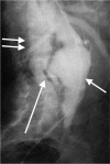
Conventional catheter aortogram demonstrating a spontaneous aorto-caval fistula (larger arrow) between AAA (smaller arrow) and the IVC (double arrow). AAA, abdominal aortic aneurysm.
Figure 8
Figure 8
Axial noncontrast CT image (A) of the thoracoabdominal aorta shows hyperdense intramural hematoma (arrow) which can mimic hyperdense crescentic sign of impending rupture of aneurysm. Axial (B) and coronal (B) CTA images shows penetrating aortic ulcer (arrow). CTA, computed tomography angiography.

References

    1. McPhee JT, Hill JS, Eslami MH. The impact of gender on presentation, therapy, and mortality of abdominal aortic aneurysm in the United States, 2001-2004. J Vasc Surg 2007;45:891-9. 10.1016/j.jvs.2007.01.043 - DOI - PubMed
    1. Pande RL, Beckman JA. Abdominal aortic aneurysm: populations at risk and how to screen. J Vasc Interv Radiol 2008;19:S2-8. 10.1016/j.jvir.2008.03.010 - DOI - PubMed
    1. Wadgaonkar AD, Black JH, 3rd, Weihe EK, et al. Abdominal Aortic Aneurysms Revisited: MDCT with Multiplanar Reconstructions for Identifying Indicators of Instability in the Pre- and Postoperative Patient. Radiographics 2015;35:254-68. 10.1148/rg.351130137 - DOI - PubMed
    1. Rakita D, Newatia A, Hines JJ, et al. Spectrum of CT findings in rupture and impending rupture of abdominal aortic aneurysms. Radiographics 2007;27:497-507. 10.1148/rg.272065026 - DOI - PubMed
    1. Rudarakanchana N, Powell JT. Advances in imaging and surveillance of AAA: when, how, how often? Prog Cardiovasc Dis 2013;56:7-12. 10.1016/j.pcad.2013.05.006 - DOI - PubMed

LinkOut - more resources