. 2017 Jul 12;3(7):e1700492.
doi: 10.1126/sciadv.1700492. eCollection 2017 Jul.

High frequency of intestinal TH17 cells correlates with microbiota alterations and disease activity in multiple sclerosis

Affiliations

High frequency of intestinal TH17 cells correlates with microbiota alterations and disease activity in multiple sclerosis

Ilaria Cosorich et al. Sci Adv. .

Abstract

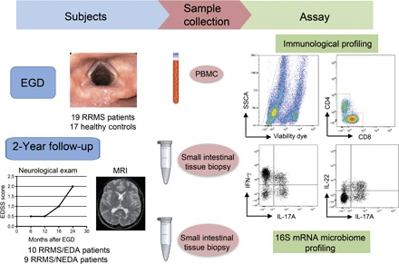
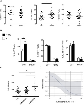
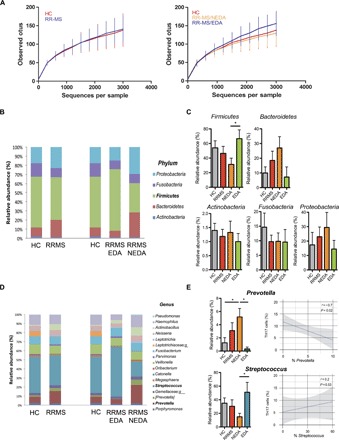
T helper 17 (TH17) cells are key players in multiple sclerosis (MS), and studies in animal models demonstrated that effector TH17 cells that trigger brain autoimmunity originate in the intestine. We validate in humans the crucial role of the intestinal environment in promoting TH17 cell expansion in MS patients. We found that increased frequency of TH17 cells correlates with high disease activity and with specific alterations of the gut mucosa-associated microbiota in MS patients. By using 16S ribosomal RNA sequencing, we analyzed the microbiota isolated from small intestinal tissues and found that MS patients with high disease activity and increased intestinal TH17 cell frequency showed a higher Firmicutes/Bacteroidetes ratio, increased relative abundance of Streptococcus, and decreased Prevotella strains compared to healthy controls and MS patients with no disease activity. We demonstrated that the intestinal TH17 cell frequency is inversely related to the relative abundance of Prevotella strains in the human small intestine. Our data demonstrate that brain autoimmunity is associated with specific microbiota modifications and excessive TH17 cell expansion in the human intestine.


Figures

Fig. 1
Fig. 1. Study design.
Tissue samples from the duodenal mucosa were collected during EGD from RRMS patients (n = 19) and HCs (n = 17). Simultaneously, we collected 6 to 7 ml of peripheral blood from the same individuals. Total lymphocytes were isolated from one to two fragments of intestinal tissue by collagenase digestion and from peripheral blood by Ficoll gradient. Mucosal immune cells and peripheral blood mononuclear cells (PBMCs) were FACS-analyzed for immunological profiling of the following TH cell subsets: IL-17A+CD4+ TH17 cells, IFN-γ+CD4+ TH1 cells, and IL-22+ TH22 cells. Microbial DNA was extracted from frozen intestinal tissue samples (seven RRMS and seven HC samples), and 16S rRNA sequencing was performed on the Roche 454 platform. At the 2-year end point, we stratified RRMS patients in two cohorts on the basis of evidence of disease activity by clinical and MRI criteria. RRMS patients with no evidence of disease activity in the 2-year follow-up were classified as RRMS/NEDA (n = 9), whereas RRMS patients with evidence of disease activity were classified as RRMS/EDA (n = 10). SSCA, side scatter FACS parameter.
Fig. 2
Fig. 2. Intestinal T cell subsets in RRMS patients and HCs.
(A) Relative percentages of CD3+, CD4+, and CD8+ cells out of total lymphocytes isolated from the small intestinal mucosa of individuals with RRMS and of HCs. (B) Frequencies of different TH cell subsets (TH17, TH1, and TH22) in the intestinal mucosa (GUT) and PBMCs of HCs and RRMS patients. Blood samples were collected from the same individuals at the time of intestinal tissue sample collection. Data are means ± SEM. (C) RRMS patients were divided into two cohorts on the basis of disease activity. RRMS patients with evidence of disease activity at 2-year follow-up were classified as EDA, whereas patients with no disease activity were classified as NEDA. Percentages of intestinal TH17 cells out of total CD4+ T cells in HCs and RRMS/NEDA and RRMS/EDA patients are shown. (A to C) **P < 0.01 and *P < 0.05 by unpaired t test. (D) Logistic regression analysis showing correlation between TH17 cell percentages in the gut mucosa and predicted probability of showing disease activity at 2-year follow-up (NEDA score: RRMS/NEDA, 1; RRMS/EDA, 0). Odds ratio, 0.96; 95% CI, 0.02 to 0.98; P = 0.01.
Fig. 3
Fig. 3. Differences of gut mucosa-associated microbiota composition between RRMS patients and HCs.
(A) α-Diversity of gut microbiota profiles assessed by 16S amplicon sequencing of DNA purified from human small intestinal tissue of HCs and RRMS patients with active (RRMS/EDA) and nonactive (RRMS/NEDA) disease. (B) Average phylum-level composition of microbiota isolated from small intestinal samples of HC and total RRMS, RRMS/NEDA, and RRMS/EDA patients. (C) Means ± SEM of the relative abundance of different phyla. *P < 0.05 by unpaired t test. (D) Average genus-level composition in four cohorts of individuals. (E) Left: Means ± SEM of the relative abundance of Prevotella and Streptococcus strains. *P < 0.05 by unpaired t test. Right: Regression linear plot in all individuals (RRMS + HC) showing inverse relation between percentages of intestinal TH17 cells and relative abundance of Prevotella strains.

References

    1. Nylander A., Hafler D. A., Multiple sclerosis. J. Clin. Invest. 122, 1180–1188 (2012). - PMC - PubMed
    1. Pette M., Fujita K., Wilkinson D., Altmann D. M., Trowsdale J., Giegerich G., Hinkkanen A., Epplen J. T., Kappos L., Wekerle H., Myelin autoreactivity in multiple sclerosis: Recognition of myelin basic protein in the context of HLA-DR2 products by T lymphocytes of multiple-sclerosis patients and healthy donors. Proc. Natl. Acad. Sci. U.S.A. 87, 7968–7972 (1990). - PMC - PubMed
    1. Odoardi F., Sie C., Streyl K., Ulaganathan V. K., Schläger C., Lodygin D., Heckelsmiller K., Nietfeld W., Ellwart J., Klinkert W. E. F., Lottaz C., Nosov M., Brinkmann V., Spang R., Lehrach H., Vingron M., Wekerle H., Flügel-Koch C., Flügel A., T cells become licensed in the lung to enter the central nervous system. Nature 488, 675–679 (2012). - PubMed
    1. Berer K., Mues M., Koutrolos M., Al Rasbi Z., Boziki M., Johner C., Wekerle H., Krishnamoorthy G., Commensal microbiota and myelin autoantigen cooperate to trigger autoimmune demyelination. Nature 479, 538–541 (2011). - PubMed
    1. Korn T., Anderson A. C., Bettelli E., Oukka M., The dynamics of effector T cells and Foxp3+ regulatory T cells in the promotion and regulation of autoimmune encephalomyelitis. J. Neuroimmunol. 191, 51–60 (2007). - PMC - PubMed

Publication types