Review
. 2017 Dec;74(24):4471-4509.
doi: 10.1007/s00018-017-2587-9. Epub 2017 Jul 13.

Epithelial cell senescence: an adaptive response to pre-carcinogenic stresses?

Affiliations
Review

Epithelial cell senescence: an adaptive response to pre-carcinogenic stresses?

Corinne Abbadie et al. Cell Mol Life Sci. 2017 Dec.

Abstract

Senescence is a cell state occurring in vitro and in vivo after successive replication cycles and/or upon exposition to various stressors. It is characterized by a strong cell cycle arrest associated with several molecular, metabolic and morphologic changes. The accumulation of senescent cells in tissues and organs with time plays a role in organismal aging and in several age-associated disorders and pathologies. Moreover, several therapeutic interventions are able to prematurely induce senescence. It is, therefore, tremendously important to characterize in-depth, the mechanisms by which senescence is induced, as well as the precise properties of senescent cells. For historical reasons, senescence is often studied with fibroblast models. Other cell types, however, much more relevant regarding the structure and function of vital organs and/or regarding pathologies, are regrettably often neglected. In this article, we will clarify what is known on senescence of epithelial cells and highlight what distinguishes it from, and what makes it like, replicative senescence of fibroblasts taken as a standard.

Keywords: Aging; Autophagy; Cancer; DNA damage; DSB; Keratinocytes; Mammary epithelial cells; Oxidative stress; PARP1; Proteostasis; SASP; SSB; Unfolded protein response; p16; p38MAPK; p53.


Conflict of interest statement

The authors declare that they have no conflict of interest.

Figures

Fig. 1
Fig. 1
Two-step senescence of human keratinocytes and mammary epithelial cells. a Schematic representation of the in vitro growth curves. b Schematic representation of DNA damage and cell cycle arrest pathways involved in the two senescence plateaus. The filled rectangles represent events common to keratinocytes and mammary epithelial cells. The hatched rectangles represent events specific to one cell type. The dashed lines represents non investigated phases
Fig. 2
Fig. 2
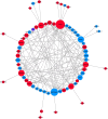
Protein–protein interactions between the known regulators of epithelial cell senescence. The 70 regulators of epithelial cell senescence listed in Table 2 plus the cell cycle regulators p53, p21, p16, RB, p15, p27, cyclin D1 and cyclin D2 were analyzed using STRING for predictive protein-protein interactions and then entered into CYTOSCAPE to visualize the network. The diameters of the circles are proportional to the number of interactions. Red circles represent positive regulators of epithelial cell senescence, blue circles negative regulators, and purple circles regulators reported either as positive or negative regulator according to the study
Fig. 3
Fig. 3
Summary of the known cellular and molecular events involved in epithelial cell senescence. Oxidative stress is at the center of the events involved in epithelial cell senescence. It is responsible, at least in part, of the DNA damages that induce the cell cycle arrest and the SASP, as well as of the induction of housekeeping machineries. It is maintained by positive loops involving some components of the SASP, the mitochondrion and the p38MAPK

References

    1. Hayflick L. The limited in vitro lifetime of human diploid cell strains. Exp Cell Res. 1965;37:614–636. doi: 10.1016/0014-4827(65)90211-9. - DOI - PubMed
    1. Hayflick L, Moorhead PS. The serial cultivation of human diploid cell strains. Exp Cell Res. 1961;25:585–621. doi: 10.1016/0014-4827(61)90192-6. - DOI - PubMed
    1. Vaziri H, Benchimol S. From telomere loss to p53 induction and activation of a DNA-damage pathway at senescence: the telomere loss/DNA damage model of cell aging. Exp Gerontol. 1996;31:295–301. doi: 10.1016/0531-5565(95)02025-X. - DOI - PubMed
    1. Hezel AF, Bardeesy N, Maser RS. Telomere induced senescence: end game signaling. Curr Mol Med. 2005;5:145–152. doi: 10.2174/1566524053586563. - DOI - PubMed
    1. Deng Y, Chan SS, Chang S. Telomere dysfunction and tumour suppression: the senescence connection. Nat Rev Cancer. 2008;8:450–458. doi: 10.1038/nrc2393. - DOI - PMC - PubMed

Publication types

LinkOut - more resources