Review
. 2017 Sep;8(3):255-272.
doi: 10.1007/s13239-017-0317-y. Epub 2017 Jul 13.

Imaging in Vascular Access

Affiliations
Review

Imaging in Vascular Access

Eoin A Murphy et al. Cardiovasc Eng Technol. 2017 Sep.

Abstract

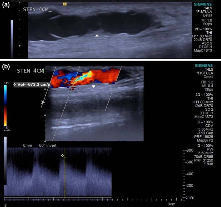
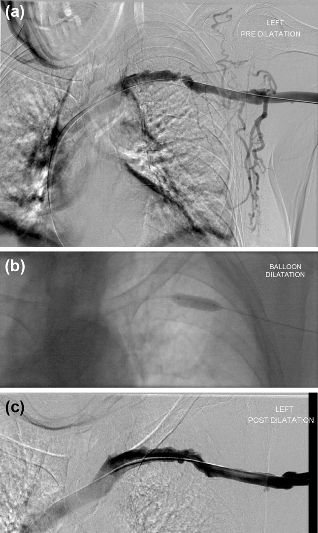
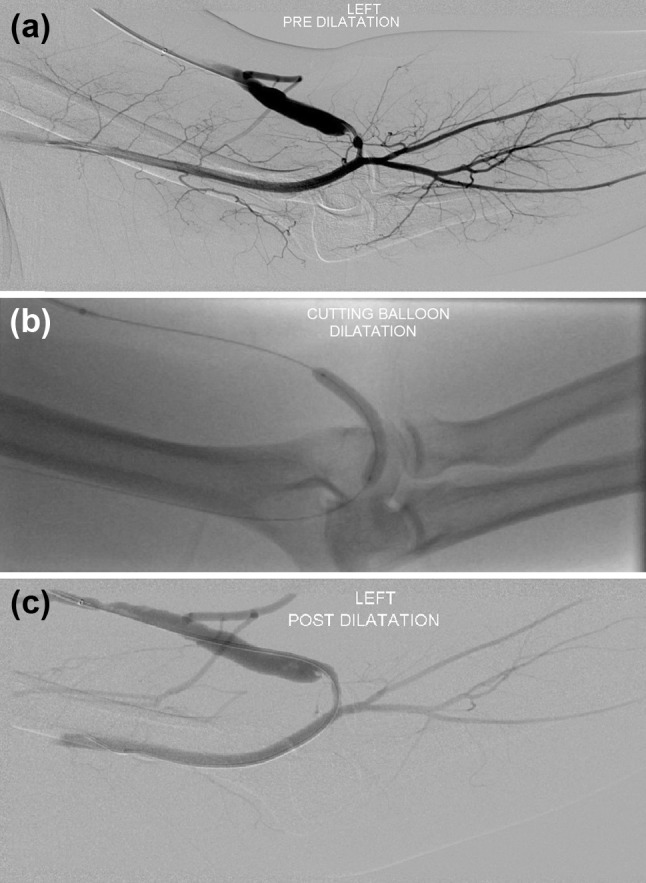
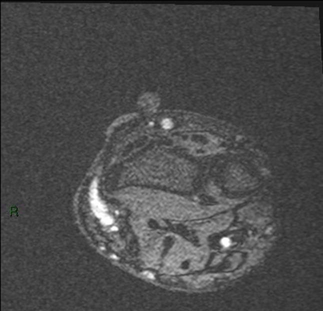
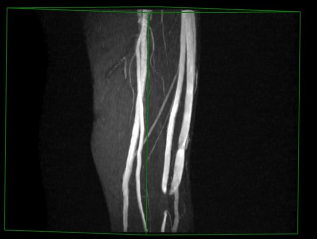
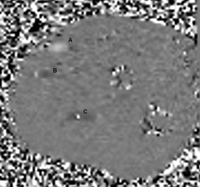
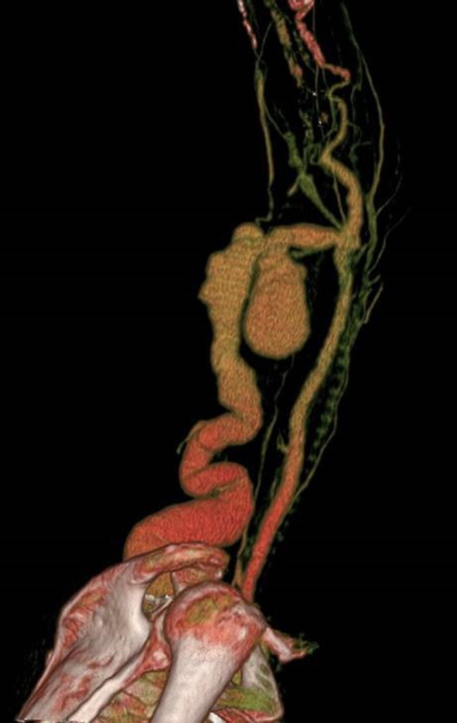
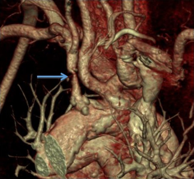
This review examines four imaging modalities; ultrasound (US), digital subtraction angiography (DSA), magnetic resonance imaging (MRI) and computed tomography (CT), that have common or potential applications in vascular access (VA). The four modalities are reviewed under their primary uses, techniques, advantages and disadvantages, and future directions that are specific to VA. Currently, US is the most commonly used modality in VA because it is cheaper (relative to other modalities), accessible, non-ionising, and does not require the use of contrast agents. DSA is predominantly only performed when an intervention is indicated. MRI is limited by its cost and the time required for image acquisition that mainly confines it to the realm of research where high resolution is required. CT's short acquisition times and high resolution make it useful as a problem-solving tool in complex cases, although accessibility can be an issue. All four imaging modalities have advantages and disadvantages that limit their use in this particular patient cohort. Current imaging in VA comprises an integrated approach with each modality providing particular uses dependent on their capabilities. MRI and CT, which currently have limited use, may have increasingly important future roles in complex cases where detailed analysis is required.

Keywords: Arteriovenous fistula; Computed tomography; Digital subtraction angiography; Magnetic resonance imaging; Medical imaging; Ultrasound; Vascular access.


Figures

Figure 1
Figure 1
(a) US b-mode image of the vein-side of an AVF with a stenosis (marked with an asterisk). Also visible are the leaflets of a venous valve fixed in position. (b) US b-mode image of the same area superimposed with the colour Doppler data, and with PSV recorded at 6.733 m/s at the location within the crosshairs (stenosis again marked with an asterisk).
Figure 2
Figure 2
(a) Left arm angiogram shows a tight stenosis of the left subclavian vein, with marked visibility of the collateral branches. (b) A 10 mm standard balloon was used for the dilatation which (c) gave a good post dilatation result.
Figure 3
Figure 3
(a) Stenosis of a left brachiocephalic fistula, just central to the anastomosis. (b) A 6 mm cutting balloon was deployed at the stenosis site. (c) Good post dilatation results were observed.
Figure 4
Figure 4
Typical TOF image showing a radiocephalic fistula swing segment as a bright region on the left. The round bright region adjacent to the arm at the top of the image is an oil capsule used as a reference marker.
Figure 5
Figure 5
A snapshot of a 3-D reconstruction of the vasculature from TOF images at the elbow region of a patient’s brachiocephalic fistula. Note the signal dropout at the anastomosis because of flow turbulence.
Figure 6
Figure 6
A snapshot of a VENC showing the radial artery as the light grey region (a) and the vein as a dark grey region (b), and some aliasing in the ulnar artery (c) because the velocities are outside the VENC range.
Figure 7
Figure 7
3D reconstruction CT scan of a left brachiocephalic AV Fistula. The patient presented with localized gross bruising and tenderness 24 h following dialysis. A pulsatile swelling was evident on clinical examination. Ultrasound examination was not possible due to patient discomfort. The image adequately demonstrates a large pseudoaneurysm arising from the proximal aspect of the AV fistula. This image provided vital pre-operative road mapping information to the surgeon before operating to repair this abnormality.
Figure 8
Figure 8
3D reconstruction CT scan of an aortic arch in a patient with a poorly maturing AV fistula. A proximal subclavian artery stenosis was identified (arrow) and subsequently treated with an endovascular stent.
Figure 9
Figure 9
(a) Maximum intensity projection (MIP) image of a left loop thigh AVG in a patient who presented with spontaneous bleeding over the AVG. It demonstrates localized breakdown of the graft material and pseudoaneurysm formation (arrow) associated with repetitive needling at the same site. This was urgently treated with insertion of a stent-graft. (b) 3D reconstruction of the same patient also shows early AVG breakdown in the medial limb of the graft (arrow).
Figure 10
Figure 10
(a) Single cone-beam CT image taken during placement of a translumbar vena cava dialysis line in a patient with no further peripheral vascular access options. The image was used to safely and accurately puncture a stenotic inferior vena cava (IVC) via the tranlumbar route. The image shows the needle is the correct position with the tip in the IVC. (b) The tunneled catheter was subsequently placed using the Seldinger technique. The tip of the catheter can be seen adequately positioned within the right atrium on this single DSA image.

References

    1. Ahmed S, Raman SP, Fishman EK. Three-dimensional MDCT angiography for the assessment of arteriovenous grafts and fistulas in hemodialysis access. Diagn. Interv. Imaging. 2016;97:297–306. doi: 10.1016/j.diii.2015.12.008. - DOI - PubMed
    1. Alley H, Owens CD, Gasper WJ, Grenon SM. Ultrasound assessment of endothelial-dependent flow-mediated vasodilation of the brachial artery in clinical research. J. Vis. Exp. 2014;92:e52070. doi: 10.3791/52070. - DOI - PMC - PubMed
    1. Allon M, Daugirdas J, Depner TA, Greene T, Ornt D, Schwab SJ. Effect of change in vascular access on patient mortality in hemodialysis patients. Am. J. Kidney Dis. 2006;47:469–477. doi: 10.1053/j.ajkd.2005.11.023. - DOI - PubMed
    1. Allon M, Lockhart ME, Lilly RZ, Gallichio MH, Young CJ, Barker J, Deierhoi MH, Robbin ML. Effect of preoperative sonographic mapping on vascular access outcomes in hemodialysis patients. Kidney Int. 2001;60:2013–2020. doi: 10.1046/j.1523-1755.2001.00031.x. - DOI - PubMed
    1. Allon M, Robbin ML. Increasing arteriovenous fistulas in hemodialysis patients: problems and solutions. Kidney Int. 2002;62:1109–1124. doi: 10.1046/j.1523-1755.2002.00551.x. - DOI - PubMed

MeSH terms