Lymphoepithelial carcinoma of salivary glands: CT and MR imaging findings
- PMID: 28707954
- PMCID: PMC5965942
- DOI: 10.1259/dmfr.20170053
Lymphoepithelial carcinoma of salivary glands: CT and MR imaging findings
Abstract
Objectives: To depict the CT and MRI characteristics of salivary gland lymphoepithelial carcinoma (LEC) and provide more diagnostic information for this malignancy.
Methods: 103 salivary gland LEC subjects were retrospectively reviewed. The subjects include 35 males with a mean age of 40.8 years and 68 females with a mean age of 49.4 years. Of the 103 subjects, 86 had carcinomas in the parotid gland, 5 in the submandibular gland, 1 in the sublingual gland, 3 in the cheek and 8 in the palate. All subjects underwent routine CT and MRI (plain and contrast-enhanced scans) prior to surgical treatment and histopathological examination.
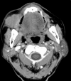
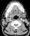
Results: Based on the pathological outcomes, all the salivary gland LECs were classified into two types from CT and MRI scans: solitary LEC (56 cases, 54.4%) and multiple LEC (47 cases, 45.6%). The latter included solitary salivary gland LEC with extraglandular lymph-node metastases (12 cases), parotid gland LEC with ipsilateral intraglandular lymph-node metastases (11 cases), parotid gland LEC with ipsilateral intra- and extraglandular lymph-node metastases (23 cases) and bilateral parotid gland LEC (1 case). The salivary gland LEC was depicted on CT and MRI scans as a lobular mass in 64 of 104 (61.5%), homogeneous mass in 65 of 104 (62.5%) or enhanced neoplasm in 94 of 104 (90.4%).
Conclusions: Salivary gland LEC has a predilection for females in the fourth to fifth decade of life and the parotid gland. CT and MRI findings between solitary and multiple salivary LECs vary. A majority of multiple parotid gland LECs are characterized by metastasis of ipsilateral intraglandular lymph nodes, which may accompany with or without extraglandular lymph-node metastases.
Keywords: computed tomography; lymphoepithelial carcinoma; magnetic resonance imaging; salivary gland.
Figures


Similar articles
-
[Analysis of the lymphoepithelial carcinoma of the salivary gland: 12 cases report].Lin Chuang Er Bi Yan Hou Tou Jing Wai Ke Za Zhi. 2017 Jul 20;31(14):1093-1096. doi: 10.13201/j.issn.1001-1781.2017.14.010. Lin Chuang Er Bi Yan Hou Tou Jing Wai Ke Za Zhi. 2017. PMID: 29798248 Chinese.
-
CT features and pathologic characteristics of lymphoepithelial carcinoma of salivary glands.Int J Clin Exp Pathol. 2014 Feb 15;7(3):1004-11. eCollection 2014. Int J Clin Exp Pathol. 2014. PMID: 24696717 Free PMC article.
-
Lymphoepithelial carcinoma of the salivary glands.Medicine (Baltimore). 2017 Feb;96(7):e6115. doi: 10.1097/MD.0000000000006115. Medicine (Baltimore). 2017. PMID: 28207533 Free PMC article.
-
Mammary analog secretory carcinoma of salivary gland with high-grade histology arising in hard palate, report of a case and review of literature.Int J Clin Exp Pathol. 2014 Dec 1;7(12):9008-22. eCollection 2014. Int J Clin Exp Pathol. 2014. PMID: 25674280 Free PMC article. Review.
-
Cervical lymph node metastasis in adenoid cystic carcinoma of the major salivary glands.J Laryngol Otol. 2017 Feb;131(2):96-105. doi: 10.1017/S0022215116009749. Epub 2016 Dec 15. J Laryngol Otol. 2017. PMID: 27974071 Review.
Cited by
-
Advantages of contrast-enhanced CT combined with DCE-MRI in identifying malignant parotid tumor.Am J Transl Res. 2022 Dec 15;14(12):9047-9056. eCollection 2022. Am J Transl Res. 2022. PMID: 36628209 Free PMC article.
-
Clinical features in salivary gland lymphoepithelial carcinoma in 10 patients: Case series and literature review.Laryngoscope Investig Otolaryngol. 2022 May 9;7(3):779-784. doi: 10.1002/lio2.811. eCollection 2022 Jun. Laryngoscope Investig Otolaryngol. 2022. PMID: 35734066 Free PMC article.
-
Prognostic Landscape of Lymphoepithelial Carcinoma: Analysis of a National Database.Cureus. 2025 Feb 10;17(2):e78828. doi: 10.7759/cureus.78828. eCollection 2025 Feb. Cureus. 2025. PMID: 40084324 Free PMC article.
-
Lymphoepithelial Carcinoma of the Sublingual Gland: A Case Report.Cureus. 2024 Feb 16;16(2):e54305. doi: 10.7759/cureus.54305. eCollection 2024 Feb. Cureus. 2024. PMID: 38496083 Free PMC article.
-
Magnetic Resonance Imaging Findings of Lymphoepithelial Carcinoma of the Submandibular Gland: A Case Report.Cureus. 2023 Dec 4;15(12):e49939. doi: 10.7759/cureus.49939. eCollection 2023 Dec. Cureus. 2023. PMID: 38179348 Free PMC article.
References
-
- Tsang WYW, Kuo TT, Chan JKC. Lymphoepithelial carcinoma :Barnes L, , Eveson JW, Reichart P, Sidransky D. World Health Organization classification of tumours. Pathology and genetics of head and neck tumours. Lyon, France: IARC Press; 2005. 251–2
-
- Tian Z, Li L, Wang L, Hu Y, Li J. Salivary gland neoplasms in oral and maxillofacial regions: a 23-year retrospective study of 6982 cases in an eastern Chinese population. Int J Oral Maxillofac Surg 2010; 39: 235–42.10.1016/j.ijom.2009.10.016 - DOI - PubMed
-
- Ban X, Wu J, Mo Y, Yang Q, Liu X, Xie C, et al. Lymphoepithelial carcinoma of the salivary gland: morphologic patterns and imaging features on CT and MRI. AJNR Am J Neuroradiol 2014; 35: 1813–9.10.3174/ajnr.A3940 - DOI - PMC - PubMed
-
- Zhan KY, Nicolli EA, Khaja SF, Day TA. Lymphoepithelial carcinoma of the major salivary glands: predictors of survival in a non-endemic region. Oral Oncol 2016; 52: 24–9.10.1016/j.oraloncology.2015.10.019 - DOI - PubMed
MeSH terms
Substances
LinkOut - more resources
Full Text Sources
Other Literature Sources
Medical

