

Encephalocraniocutaneous lipomatosis: A case report with review of literature
- PMID: 28707961
- PMCID: PMC5703133
- DOI: 10.1177/1971400917693638
Encephalocraniocutaneous lipomatosis: A case report with review of literature
Abstract
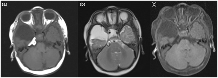
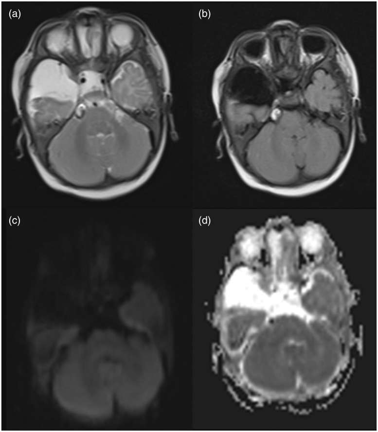
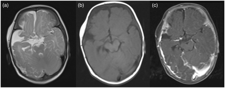
Encephalocraniocutaneous lipomatosis (ECCL) or Haberland syndrome is an uncommon sporadic neurocutaneous syndrome of unknown origin. The rarity and common ignorance of the condition often makes diagnosis difficult. The hallmark of this syndrome is the triad of skin, ocular and central nervous system (CNS) involvement and includes a long list of combination of conditions. Herein we report a case of a 5-month-old male child who presented to our centre with complaint of seizure. The patient had various cutaneous and ocular stigmatas of the disease in the form of patchy alopecia of the scalp, right-sided limbal dermoid and a nodular skin tag near the lateral canthus of the right eye. MRI of the brain was conducted which revealed intracranial lipoma and arachnoid cyst. The constellation of signs and symptoms along with the skin, ocular and CNS findings led to the diagnosis of ECCL.
Keywords: Encephalocraniocutaneous lipomatosis; Haberland syndrome; neurocutaneous syndrome.
Figures
Similar articles
-
Encephalocraniocutaneous Lipomatosis: Haberland Syndrome.Am J Case Rep. 2017 Dec 1;18:1271-1275. doi: 10.12659/ajcr.907685. Am J Case Rep. 2017. PMID: 29192135 Free PMC article.
-
Encephalocraniocutaneous lipomatosis (ECCL): neuroradiological findings in three patients and a new association with fibrous dysplasia.Am J Med Genet A. 2011 Jul;155A(7):1690-6. doi: 10.1002/ajmg.a.33954. Epub 2011 May 27. Am J Med Genet A. 2011. PMID: 21626669 Review.
-
Bilateral ocular involvement in encephalocraniocutaneous lipomatosis.Eur J Paediatr Neurol. 2007 Mar;11(2):108-10. doi: 10.1016/j.ejpn.2006.11.002. Epub 2007 Jan 26. Eur J Paediatr Neurol. 2007. PMID: 17258481
-
Encephalocraniocutaneous lipomatosis: a case report and review of the literature.Indian J Ophthalmol. 2014 May;62(5):622-7. doi: 10.4103/0301-4738.133521. Indian J Ophthalmol. 2014. PMID: 24881613 Free PMC article. Review.
-
Lipomatosis encefalocraneocutánea: reporte de caso.Gac Med Mex. 2017;153(7):915-918. doi: 10.24875/GMM.17002833. Gac Med Mex. 2017. PMID: 29414957
Cited by
-
Ophthalmic Manifestation and Pathological Features in a Cohort of Patients With Linear Nevus Sebaceous Syndrome and Encephalocraniocutaneous Lipomatosis.Front Pediatr. 2021 May 20;9:678296. doi: 10.3389/fped.2021.678296. eCollection 2021. Front Pediatr. 2021. PMID: 34095036 Free PMC article.
-
Encephalocraniocutaneous Lipomatosis: Haberland Syndrome.Am J Case Rep. 2017 Dec 1;18:1271-1275. doi: 10.12659/ajcr.907685. Am J Case Rep. 2017. PMID: 29192135 Free PMC article.
-
Encephalocraniocutaneous Lipomatosis, a Radiological Challenge: Two Atypical Case Reports and Literature Review.Brain Sci. 2022 Nov 30;12(12):1641. doi: 10.3390/brainsci12121641. Brain Sci. 2022. PMID: 36552101 Free PMC article.
-
Update of Pediatric Lipomatous Lesions: A Clinicopathological, Immunohistochemical and Molecular Overview.J Clin Med. 2022 Mar 31;11(7):1938. doi: 10.3390/jcm11071938. J Clin Med. 2022. PMID: 35407546 Free PMC article. Review.
References
-
- Haberland C, Perou M. Encephalocraniocutaneous lipomatosis: A new example of ectomesodermal dysgenesis. Arch Neurol 1970; 22: 144–155. - PubMed
-
- Fishman MA, Chang CS, Miller JE. Encephalocraniocutaneous lipomatosis. Pediatrics 1978; 61: 580–582. - PubMed
-
- Happle R, Kuster W. Nevus psiloliparus: A distinct fatty tissue nevus. Dermatology 1998; 197: 6–10. - PubMed
-
- Parazzini C, Triulzi F, Russo G, et al. Encephalocraniocutaneous lipomatosis: Complete neuroradiologic evaluation and follow-up of two cases. AJNR Am J Neuroradiol 1999; 20: 173–176. - PubMed
-
- Hunter AG. Oculocerebrocutaneous and encephalocraniocutaneous lipomatosis syndromes: Blind men and an elephant or separate syndromes? Am J Med Genet A 2006; 140: 709–726. - PubMed
Publication types
MeSH terms
Substances
Supplementary concepts
LinkOut - more resources
Full Text Sources
Other Literature Sources
Medical
