. 2017 Nov;24(11):1861-1871.
doi: 10.1038/cdd.2017.106. Epub 2017 Jul 14.

Regulation of PLK1 through competition between hnRNPK, miR-149-3p and miR-193b-5p

Affiliations

Regulation of PLK1 through competition between hnRNPK, miR-149-3p and miR-193b-5p

Chang Hoon Shin et al. Cell Death Differ. 2017 Nov.

Abstract

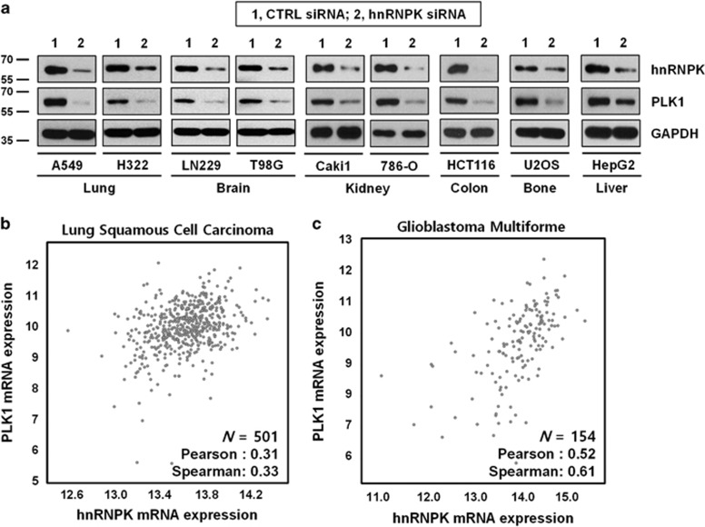
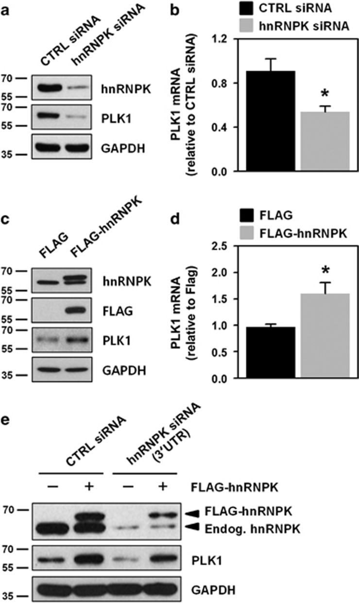
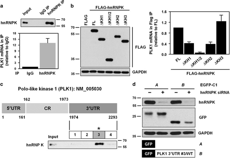
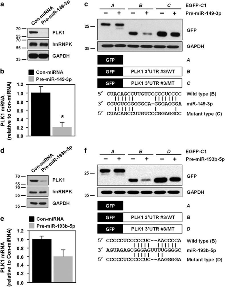
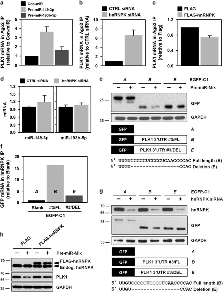
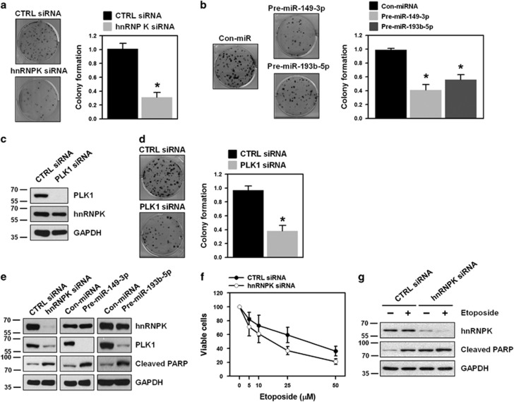
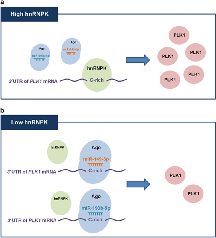
Polo-like kinase 1 (PLK1) is a critical regulator of cell cycle progression and apoptosis. However, its regulation remains poorly understood. In the present study, we investigated the molecular mechanism underlying the post-transcriptional regulation of PLK1. We observed that heterogeneous nuclear ribonucleoprotein K (hnRNPK) and PLK1 were positively associated in several different cancers and high expression levels of them correlated with poor prognosis in patients with cancer. Knockdown of hnRNPK resulted in reduced expression of PLK1, whereas conversely, PLK1 expression was increased in hnRNPK-overexpressing cells. We found that hnRNPK regulated PLK1 expression through KH1- and KH2-dependent interactions with the 3'UTR of PLK1 mRNA. In addition, microRNA-149-3p (miR-149-3p) and miR-193b-5p suppressed PLK1 expression by targeting the 3'UTR of PLK1 mRNA. MicroRNA-elicited enrichment of PLK1 mRNA in Ago2 immunoprecipitation was altered by the presence or absence of hnRNPK. Furthermore, the deletion of the cytosine (C)-rich sequences of the 3'UTR of PLK1 mRNA abolished the decreased PLK1 expression observed via hnRNPK silencing and administration of miRNAs, a finding that suggests that hnRNPK shares this C-rich motif with miR-149-3p and miR-193b-5p. We also found that downregulation of PLK1 by either silencing hnRNPK or overexpression of miR-149-3p and miR-193b-5p decreased clonogenicity and induced apoptosis. Our findings from this study demonstrate that hnRNPK regulates PLK1 expression by competing with the PLK1-targeting miRNAs, miR-149-3p and miR-193b-5p.


Conflict of interest statement

The authors declare no conflict of interest.

Figures

Figure 1
Figure 1
Positive correlation between hnRNPK and PLK1 in several types of cancer cells. (a) To examine whether hnRNPK is involved in PLK1 expression, cells from the following cancer cell lines were transfected with hnRNPK-specific siRNA: lung adenocarcinoma (A549 and H322), glioblastoma (LN229 and T98G), renal cell carcinoma (Caki1 and 786-O), colon cancer (HCT116), osteosarcoma (U2OS), and hepatocellular carcinoma (HepG2). The level of hnRNPK and PLK1 was determined by western blot. As a loading control, the level of GAPDH was examined. (b,c) To validate positive correlations, the mRNA level of hnRNPK and PLK1 in lung squamous cell carcinoma (b) and glioblastoma (c) was obtained from the TCGA database
Figure 2
Figure 2
hnRNPK regulates PLK1 expression. (a,b) HeLa cells were transfected with control or hnRNPK-specific siRNA. After 48 h post-transfection, protein and mRNA levels of hnRNPK and PLK1 were determined by western blot and RT–qPCR, respectively. (c,d) Cells were transfected with FLAG or FLAG-hnRNPK vector. Protein and mRNA levels of hnRNPK and PLK1 were assessed as described above. (e) To verify that hnRNPK regulates PLK1, we designed a specific siRNA targeting the 3′UTR of hnRNPK mRNA (Supplementary Table S2). HeLa cells were simultaneously transfected with siRNAs (control or hnRNPK 3′UTR siRNA) and with plasmid DNA (FLAG or FLAG-hnRNPK vector). After 48 h post-transfection, the level of endogenous and ectopic hnRNPK, PLK1, and the loading control, GAPDH, was assessed by western blot. All experiments were performed more than three times and data represent mean±S.D.
Figure 3
Figure 3
hnRNPK directly interacts with the 3′UTR of PLK1 mRNA through its KH1 and KH2 domains. (a) To determine whether hnRNPK binds to the 3′UTR of PLK1 mRNA, cytoplasmic lysates were prepared as described in the Materials and Methods, and immunoprecipitated (IP) using hnRNPK antibody-coated beads. RNA was isolated from IP materials and the level of PLK1 mRNA was determined by RT–qPCR. Western blot was performed to confirm IP efficiency. (b) As an RNA-binding protein, hnRNPK has three KH domains: KH1, KH2, and KH3. To evaluate which domain or domains are responsible for the interaction between hnRNPK and PLK1 mRNA, four KH deletion mutants were manufactured: ΔKH1, ΔKH1/2, ΔKH2, and ΔKH3 (Supplementary Figure S3). HeLa cells were transfected with FLAG vectors containing the wild-type (full length, FL) and the four deletion mutants. Cytoplasmic lysates were prepared and IP was performed using FLAG antibody. Identical expression levels of ectopic hnRNPK (FLAG-hnRNPK) in input were determined by western blot, and the level of enriched PLK1 mRNA in FLAG-IP was assessed by RT–qPCR. (c) Schematic depiction of the biotinylated RNAs of the 3′UTR of PLK1 mRNA used for biotin pull-down analysis. The level of hnRNPK in biotin pull-down samples was determined by western blot. (d) We constructed vectors expressing chimeric RNAs spanning the GFP (A) and fragment #3 of the 3′UTR PLK1 mRNA (B). HeLa cells were first transfected with control or hnRNPK siRNA. After 24 h post-transfection, cells were resuspended into six-well plates followed by transfection with GFP vectors (blank or fragment #3 of the 3′UTR PLK1 mRNA). The level of hnRNPK, GFP, and the loading control, GAPDH, was assessed by western blot. All experiments were performed more than three times and data represent mean±S.D.
Figure 4
Figure 4
miR-149-3p and miR-193b-5p suppress PLK1 expression through direct interactions with the 3′UTR of PLK1 mRNA. (a,b) HeLa cells were transfected with control miRNA or pre-miR-149-3p. After 48 h post-transfection, PLK1 protein and mRNA levels were determined by western blot and RT–qPCR, respectively. hnRNPK and the loading control, GAPHD, were examined by western blot. (c) We constructed two GFP vectors to investigate whether a direct interaction with the 3′UTR PLK1 mRNA is required for downregulation of PLK1 by miR-149-3p. Based on vectors expressing chimeric RNAs spanning the GFP (A) and fragment #3 of the 3′UTR PLK1 mRNA (B), we constructed vectors (C) containing mutated sequences of the miR-149-3p-binding sites in the 3′UTR PLK1 mRNA (fragment #3). After overexpression of miR-149-3p, cells were transfected with the indicated GFP vectors: blank, wild-type (WT), or mutated type (MT). (df) Similar to our investigation of miR-149-3p, we tested the effect of miR-193b-5p on PLK1 expression as described above. All experiments were performed more than three times and data represent mean±S.D.
Figure 5
Figure 5
Competitive regulation of PLK1 by hnRNPK and miR-149-3p/193b-5p. (a) Cytoplasmic lysates were obtained from miR-149-3p- or miR-193b-5p-overexpressing cells and immunoprecipitated (IP) with Ago2-specific antibody. Enrichment of PLK1 mRNA in Ago2 IP was assessed by RT–qPCR. (b,c) To investigate the effect of hnRNPK on the interaction between PLK1 mRNA and an miRNA-loaded RISC complex, enrichment of PLK1 mRNA in Ago2 IP was examined using cytoplasmic lysates obtained from hnRNPK-silenced (b) or -overexpressing (c) HeLa cells. The level of PLK1 mRNA in Ago2 IP was assessed by RT–qPCR. (d) To test whether hnRNPK affects expression of miR-149-3p and miR-193b-5p, cells were transfected with control or hnRNPK siRNA. After 48 h post-transfection, miRNA expression levels were determined by RT–qPCR. (e) To examine the effect of miRNA mimics on GFP expression, GFP reporters were generated in which GFP was linked to fragment #3 harboring or lacking the binding sequence for hnRNPK and the miRNAs (GFP vector B and E in the schematic). GFP expression was assessed by western blot. (f) The interaction between hnRNPK and GFP chimeric mRNAs was examined. Cells were transfected with the previously described GFP vectors and cytoplasmic lysates were prepared. GFP mRNA enrichment was measured by RNP IP using hnRNPK antibody followed by RT–qPCR. (g) To test whether hnRNPK affects GFP expression in the absence of an miRNA-binding sequence, GFP reporters described in e were used. The expression levels of hnRNPK and GFP were assessed by western blot. (h) To determine whether hnRNPK restored PLK1 expression, cells were transfected with miRNA mimics (pre-miR-149-3p and miR-193b-5p) and an hnRNPK vector (FLAG-hnRNPK). Expression of hnRNPK and PLK1 was assessed by western blot. All experiments were performed more than three times and data represent mean±S.D.
Figure 6
Figure 6
PLK1 regulation through interplay between hnRNPK and miRNAs is implicated in clonogenicity and drug resistance. HeLa cells were transfected with hnRNPK siRNA (a), miRNA mimics (pre-miR-149-3p and miR-193b-5p) (b), or PLK1 siRNA (cd). After 48 h post-transfection, cells were resuspended into six-well plates and cultured for 2 weeks. Representative images are shown and clonogenic abilities were determined by counting the number of colonies. (e) To determine whether downregulation of PLK1 induces apoptotic cell death, HeLa cells were transfected with hnRNPK siRNA or miRNA mimics (pre-miR-149-3p and miR-193b-5p). The level of hnRNPK, PLK1, and cleaved PARP was assessed by western blot. (f) HeLa cells were transfected with control or hnRNPK-specific siRNA. Transfected cells were resuspended into 96-well plates and treated with different concentrations of etoposide. After incubation for 48 h, cell viability was determined by the MTS cell proliferation assay. (g) To test whether knockdown of hnRNPK sensitized cancer cells to etoposide treatment, HeLa cells were transfected with control or hnRNPK siRNA. Transfected cells were resuspended into 96-well plates and treated with 10 μM etoposide for 48 h. The level of hnRNPK and cleaved PARP was assessed by western blot. All experiments were performed more than three times and data represent mean±S.D.
Figure 7
Figure 7
Proposed action mechanism underlying hnRNPK-mediated PLK1 regulation. Under the condition of high hnRNPK, the interaction between PLK1 mRNA 3′UTR and miRNA-loaded RISC is disrupted by hnRNPK, which results in increase of PLK1 expression. Conversely, in the presence of low hnRNPK, miRNA-loaded RISC easily interacts with the 3′UTR of PLK1 mRNA, in turn lowering the PLK1 expression

Similar articles

Cited by

References

    1. Hamanaka R, Maloid S, Smith MR, O'Connell CD, Longo DL, Ferris DK. Cloning and characterization of human and murine homologues of the Drosophila polo serine-threonine kinase. Cell growth differ 1994; 5: 249–257. - PubMed
    1. Clay FJ, McEwen SJ, Bertoncello I, Wilks AF, Dunn AR. Identification and cloning of a protein kinase-encoding mouse gene, Plk, related to the polo gene of Drosophila. Proc Natl Acad Sci USA 1993; 90: 4882–4886. - PMC - PubMed
    1. Lake RJ, Jelinek WR. Cell cycle- and terminal differentiation-associated regulation of the mouse mRNA encoding a conserved mitotic protein kinase. Mol cell biol 1993; 13: 7793–7801. - PMC - PubMed
    1. Simmons DL, Neel BG, Stevens R, Evett G, Erikson RL. Identification of an early-growth-response gene encoding a novel putative protein kinase. Mol cell biol 1992; 12: 4164–4169. - PMC - PubMed
    1. Takai N, Hamanaka R, Yoshimatsu J, Miyakawa I. Polo-like kinases (Plks) and cancer. Oncogene 2005; 24: 287–291. - PubMed

MeSH terms

LinkOut - more resources