. 2017 Oct;24(10):1811-1820.
doi: 10.1038/cdd.2017.121. Epub 2017 Jul 14.

Blocking endothelin-1-receptor/β-catenin circuit sensitizes to chemotherapy in colorectal cancer

Affiliations

Blocking endothelin-1-receptor/β-catenin circuit sensitizes to chemotherapy in colorectal cancer

Roberta Cianfrocca et al. Cell Death Differ. 2017 Oct.

Abstract

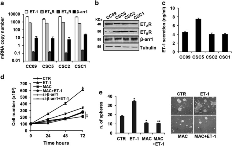
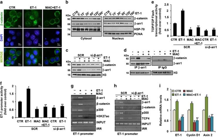
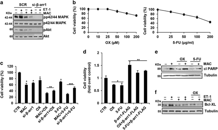
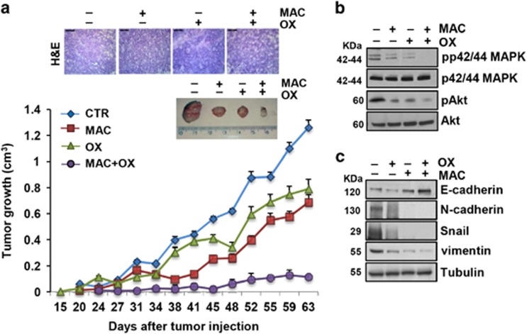
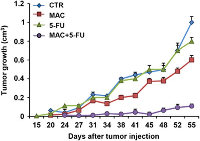
The limited clinical response to conventional chemotherapeutics observed in colorectal cancer (CRC) may be related to the connections between the hyperactivated β-catenin signaling and other pathways in CRC stem-like cells (CRC-SC). Here, we show the mechanistic link between the endothelin-1 (ET-1)/ET-1 receptor (ET-1R) signaling and β-catenin pathway through the specific interaction with the signal transducer β-arrestin1 (β-arr1), which initiates signaling cascades as part of the signaling complex. Using a panel of patient-derived CRC-SC, we show that these cells secrete ET-1 and express ETAR and β-arr1, and that the activation of ETAR/β-arr1 axis promotes the cross-talk with β-catenin signaling to sustain stemness, epithelial-to-mesenchymal transition (EMT) phenotype and response to chemotherapy. Upon ETAR activation, β-arr1 acts as a transcription co-activator that binds β-catenin, thereby promoting nuclear complex with β-catenin/TFC4 and p300 and histone acetylation, inducing chromatin reorganization on target genes, such as ET-1. The enhanced transcription of ET-1 increases the self-sustained ET-1/β-catenin network. All these findings provide a strong rationale for targeting ET-1R to hamper downstream β-catenin/ET-1 autocrine circuit. Interestingly, treatment with macitentan, a dual ETAR and ETBR antagonist, able to interfere with tumor and microenvironment, disrupts the ET-1R/β-arr1-β-catenin interaction impairing pathways involved in cell survival, EMT, invasion, and enhancing sensitivity to oxaliplatin (OX) and 5-fluorouracil (5-FU). In CRC-SC xenografts, the combination of macitentan and OX or 5-FU enhances the therapeutic effects of cytotoxic drugs. Together, these results provide mechanistic insight into how ET-1R coopts β-catenin signaling and offer a novel therapeutic strategy to manage CRC based on the combination of macitentan and chemotherapy that might benefit patients whose tumors show high ETAR and β-catenin expression.


Conflict of interest statement

The authors declare no conflict of interest.

Figures

Figure 1
Figure 1
Expression of ET-1/β-arr1 axis in CRC-SC. (a) In a panel of patient-derived CRC-SC (CC09, CSC5, CSC2 and CSC1), expression of ET-1, ETAR, ETBR and β-arr1 mRNA copy number was analyzed by real-time PCR (qPCR) normalized using endogenous cyclophilin-A. Values are shown as mean±S.D. from three independent experiments repeated in triplicates. (b) ETAR, ETBR and β-arr1 protein expression was analyzed by immunoblotting (IB). Tubulin was used as loading control. (c) ET-1 secretion evaluated by ELISA in 24 h cell-conditioned media. Values are shown as mean±S.D. from three independent experiments repeated in triplicates. (d) Time-dependent effect of treatment with ET-1 (100 nM) and/or macitentan (MAC) (1 μM) on cell growth of CC09 cells transfected with siRNA negative control (SCR) or with β-arr1 siRNA (si-β-arr1). Values are shown as mean±S.D. from three independent experiments repeated in triplicates (*P<0.001 versus CTR; **P<0.001 versus ET-1) (e) Sphere formation assay of CSC5 cells, treated with ET-1 (100 nM) and MAC (1 μM) alone or in combination for 7 days. Values are shown as mean±S.D. from three independent experiments repeated in triplicates (*P<0.05 versus CTR; **P<0.05 versus ET-1). Representative images of tumor spheres were shown in the right panel
Figure 2
Figure 2
ET-1R/β-arr1 axis drives EMT process in CRC-SC. (a) Lysates from CC09 cells were analyzed by IB for the expression of epithelial (E-cadherin) and mesenchymal (N-cadherin, Snail, and vimentin) markers after ET-1 (100 nM) stimulation for 24 h. Tubulin was used as loading control. (b) Lysates from CC09 cells transfected with SCR or si-β-arr1 and treated for 24 h with ET-1 (100 nM) and/or MAC (1 μM) were analyzed by IB for the expression of E-cadherin, N-cadherin Snail and Twist. Tubulin was used as loading control. (c) Snail, E-cadherin, and N-cadherin expression in CC09 cells upon ET-1 (100 nM) and/or MAC (1 μM) treatment evaluated by qPCR, normalized using endogenous cyclophilin-A. Values are shown as mean±S.D. from three independent experiments repeated in triplicates (*P<0.002 versus CTR; **P<0.005 versus ET-1). (d) E-cadherin promoter activity and Snail promoter activity (e) evaluated in CC09 cells transfected with SCR or si-β-arr1 and treated for 24 h with ET-1(100 nM) and/or MAC (1 μM). Values are shown as mean±S.D. from three independent experiments repeated in triplicates (*P<0.01 versus CTR; **P<0.001 versus ET-1 in SCR-transfected cells). (f) Lysates from CC09 cells transfected with SCR or si-β-arr1 treated with ET-1 (100 nM) and/or MAC (1 μM) for 24 h were analyzed for MMP-2 and -9 by IB. Tubulin was used as loading control. (g) Conditioned media collected from CCO9 treated as in (f) were used to determine the secretion and activity of MMP-2 and -9 by gelatin zymography. (h) Cell invasion assay of CC09 cells transfected with SCR or si-β-arr1 and exposed to ET-1 (100 nM) and/or MAC(1 μM) for 24 h. Values are shown as mean±S.D. from three independent experiments repeated in triplicates (*P<0.002 versus CTR; **P<0.001 versus ET-1 in SCR-transfected cells). Representative images of invading cells were shown in the right panel
Figure 3
Figure 3
ET-1R/β-arr1 axis induces β-catenin pathway in CRC-SC. (a) β-catenin localization evaluated by immunofluorescence staining (green) in CC09 cells stimulated for 30 min with ET-1 (100 nM) alone or in combination with MAC (1 μM). Nuclei were counterstained with DAPI (blue) (scale bar, 10 μm). (b) IB analysis for β-catenin and β-arr1 protein expression in cytoplasmic and nuclear extract of CC09 cells treated with ET-1(100 nM) for the indicated times. HSP-70 and PCNA were used as cytoplasmic and nuclear loading control, respectively. (c) IB analysis for β-catenin and β-arr1 protein expression in nuclear extracts of CC09 cells transfected with SCR or si-β-arr1 and treated for 30 min with ET-1 (100 nM) and/or MAC (1 μM). Histone H3 (H3) was used as loading control. (d) Nuclear extracts of CC09 cells, treated for 30 min with ET-1 (100 nM) and/or MAC (1 μM), were immunoprecipitated (IP) with anti-β-arr1 or with irrelevant IgG (IgG) and immunoblotted with anti-β-catenin and anti-β-arr1. H3 was used as loading control. (e) β-catenin transcriptional activity evaluated in CC09 cells transfected with SCR or si-β-arr1 and treated for 24 h with ET-1 (100 nM) and/or MAC (1 μM). Values are shown as mean±S.D. from three independent experiments repeated in triplicates (*P<0.001 versus CTR; **P<0.002 versus ET-1 in SCR-transfected cells). (f) ET-1 promoter activity evaluated in CC09 cells treated as in (e). Values are shown as mean±S.D. from three independent experiments repeated in triplicates (*P<0.001 versus CTR; **P<0.001 versus ET-1 in SCR-transfected cells). ChIP analysis performed in CC09 cells treated for 30 min with ET-1 (100 nM) and/or MAC (1 μM) (g) or in CC09 cells transfected with SCR or si-β-arr1 and treated for 30 min with ET-1 (100 nM) and/or MAC (1 μM) (h). Chromatin was incubated with β-arr1, β-catenin, p300, H3K27ac and TCF4 Abs and analyzed by PCR analysis by using specific primers for ET-1 promoter. Non-specific immunoglobulin G (IgG) was used as irrelevant Ab (IRR). The input DNA lane represents one-twentieth of the precleared chromatin used in each ChIP reaction. (i) ET-1, Cyclin D1 and Axin 2 mRNA expression in CC09 cells stimulated for 24 h with ET-1 (100 nM) alone or in combination with MAC (1 μM) evaluated by qPCR, normalized using endogenous cyclophilin-A. Values are shown as mean±S.D. from three independent experiments repeated in triplicates (*P<0.01 versus CTR; **P<0.05 versus ET-1)
Figure 4
Figure 4
ET-1R blockade sensitizes CRC-SC to standard chemotherapeutic drugs. (a) Lysates from CC09 cells treated for 1 h with ET-1 (100 nM) and/or MAC (1 μM) and transfected with SCR or with si-β-arr1 were immunoblotted with anti-pp42/44MAPK, anti-p42/44MAPK, anti-pAkt and anti-Akt. (b) Effect of exposure to different concentrations of oxaliplatin (OX) and 5-fluorouracil (5-FU) after 24 h on cell vitality of CC09 cells. (c) Time-dependent effect of treatment with MAC (1 μM) or OX (100 μM) or 5-FU (50 μg/ml) alone and combination, for 24 h on cell growth of CC09 cells transfected with SCR or with si-β-arr1. Values are shown as mean±S.D. from three independent experiments repeated in triplicates (*P<0.001 versus CTR; **P<0.001 versus OX; ***P<0.002 versus 5-FU). (d) Effect of treatment with OX (100 μM) or 5-FU (50 μg/ml), for 24 h on cell growth of CC09 cells transfected with empty vector (Mock) or with β-arr1-FLAG. Values are shown as mean±S.D. from three independent experiments repeated in triplicates (*P<0.002 versus CTR; **P<0.002 versus chemotherapy-treated cells). (e) IB analysis of PARP cleaved form (cl PARP) in CC09 cells treated for 24 h with MAC (1 μM) or OX (100 μM) or 5-FU (50 μg/ml) alone and combination. Tubulin was used as loading control. (f) IB analysis of Bcl-XL in CC09 cells treated for 24 h with ET-1 (100 nM) or MAC (1 μM) or OX (100 μM) alone and combination. Tubulin was used as loading control
Figure 5
Figure 5
ET-1R blockade by macitentan inhibits tumor growth and restores sensitivity to oxaliplatin in CRC-SC xenografts. (a) CC09 cells (5 × 10⁵) were injected s.c. into the flank of nude mice. When tumors were detected, mice were treated with vehicle (CTR), or MAC (30 mg/Kg/oral daily), or OX (0.25 mg/Kg/i.p. once a week), or MAC (30 mg/Kg/oral daily) with OX (0.25 mg/Kg/i.p. once a week) combination for 4 weeks. The comparison of the time course of tumor growth curves by two-way ANOVA with group-by-time interaction for tumor growth was statistically significant (P<0.02). Data points, averages±S.D. The upper panels represented the hematoxylin–eosin staining of transplanted tumor xenografts (scale bar, 50 μm) or the images of tumors from each treatment group. (b) Expression of pp42/44MAPK, p42/44MAPK, pAkt and Akt as evaluated by IB on total extracts from tumors of CC09 xenografts. (c) E-cadherin, N-cadherin, Snail and vimentin, evaluated by IB of total extracts from tumors of CC09 xenografts
Figure 6
Figure 6
Macitentan inhibits tumor growth and restores sensitivity to 5-FU in CRC-SC xenografts. CC09 cells (5 × 10⁵) were injected s.c. into the flank of nude mice. When tumors were detected, mice were treated with vehicle (CTR), or MAC (30 mg/Kg/oral daily), or 5-FU (15 mg/Kg/i.p daily), or MAC (30 mg/Kg/oral daily) with 5-FU (15 mg/Kg/i.p daily) combination for 4 weeks. The comparison of the time course of tumor growth curves by two-way ANOVA with group-by-time interaction for tumor growth was statistically significant (P<0.05). Data points, averages±S.D.

References

    1. Walker AS, Johnson EK, Maykel JA, Stojadinovic A, Nissan A, Brucher B et al. Future directions for the early detection of colorectal cancer recurrence. J Cancer 2014; 5: 272–280. - PMC - PubMed
    1. Clevers H. Wnt/beta-catenin signaling in development and disease. Cell 2006; 127: 469–480. - PubMed
    1. De Angelis ML, Zeuner A, Policicchio E, Russo G, Bruselles A, Signore M et al. Cancer stem cell-based models of colorectal cancer reveal molecular determinants of therapy resistance. Stem Cell Transl Med 2016; 5: 511–523. - PMC - PubMed
    1. Zeuner A, Todaro M, Stassi G, De Maria R. Colorectal cancer stem cells: from the crypt to the clinic. Cell Stem Cell 2014; 15: 692–705. - PubMed
    1. Colak S, Medema JP. Cancer stem cells-important players in tumor therapy resistance. FEBS J 2014; 281: 4779–4791. - PubMed

MeSH terms