

SonoNet: Real-Time Detection and Localisation of Fetal Standard Scan Planes in Freehand Ultrasound
- PMID: 28708546
- PMCID: PMC6051487
- DOI: 10.1109/TMI.2017.2712367
SonoNet: Real-Time Detection and Localisation of Fetal Standard Scan Planes in Freehand Ultrasound
Abstract
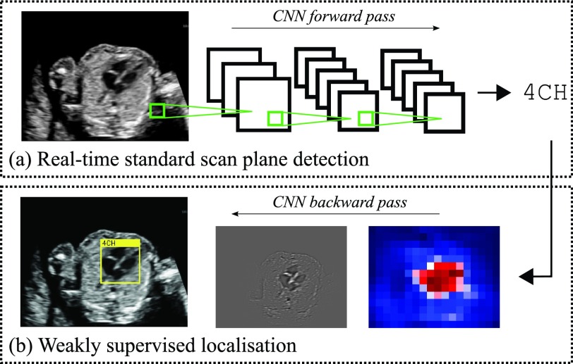
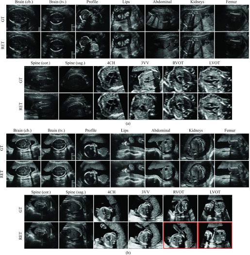
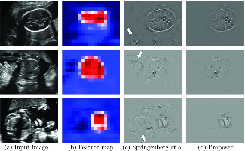
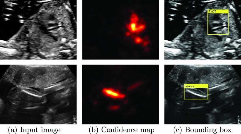
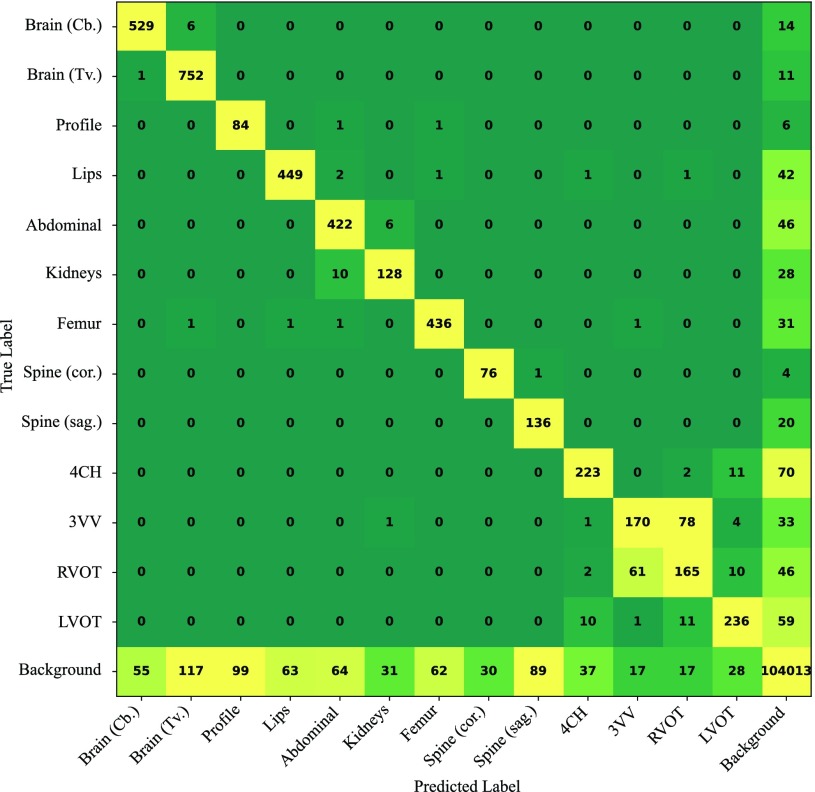
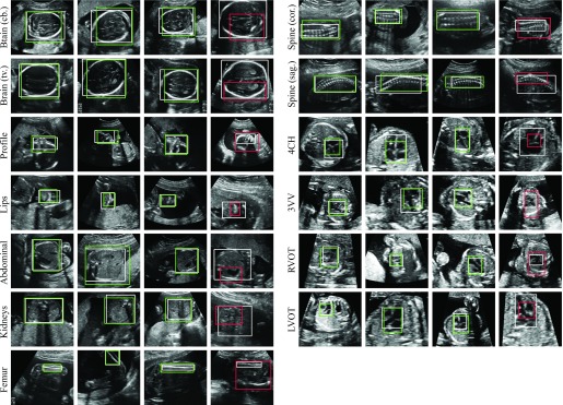
Identifying and interpreting fetal standard scan planes during 2-D ultrasound mid-pregnancy examinations are highly complex tasks, which require years of training. Apart from guiding the probe to the correct location, it can be equally difficult for a non-expert to identify relevant structures within the image. Automatic image processing can provide tools to help experienced as well as inexperienced operators with these tasks. In this paper, we propose a novel method based on convolutional neural networks, which can automatically detect 13 fetal standard views in freehand 2-D ultrasound data as well as provide a localization of the fetal structures via a bounding box. An important contribution is that the network learns to localize the target anatomy using weak supervision based on image-level labels only. The network architecture is designed to operate in real-time while providing optimal output for the localization task. We present results for real-time annotation, retrospective frame retrieval from saved videos, and localization on a very large and challenging dataset consisting of images and video recordings of full clinical anomaly screenings. We found that the proposed method achieved an average F1-score of 0.798 in a realistic classification experiment modeling real-time detection, and obtained a 90.09% accuracy for retrospective frame retrieval. Moreover, an accuracy of 77.8% was achieved on the localization task.
Figures
References
-
- Abuhamad A., Falkensammer P., Reichartseder F., and Zhao Y., “Automated retrieval of standard diagnostic fetal cardiac ultrasound planes in the second trimester of pregnancy: A prospective evaluation of software,” Ultrasound Obstetrics, Gynecol., vol. 31, no. 1, pp. 30–36, 2008. - PubMed
-
- Baumgartner C. F., Kamnitsas K., Matthew J., Smith S., Kainz B., and Rueckert D., “Real-time standard scan plane detection and localisation in fetal ultrasound using fully convolutional neural networks,” in Medical Image Computing and Computer-Assisted Intervention—MICCAI. Cham, Switzerland: Springer, 2016, pp. 203–211.
-
- Boykov Y., Veksler O., and Zabih R., “Fast approximate energy minimization via graph cuts,” IEEE Trans. Pattern Anal. Mach. Intell., vol. 23, no. 11, pp. 1222–1239, Nov. 2001.
-
- Bridge C. P., Ioannou C., and Noble J. A., “Automated annotation and quantitative description of ultrasound videos of the fetal heart,” Med. Image Anal., vol. 36, pp. 147–161, Feb. 2017. - PubMed
-
- Bridge C. P. and Noble J. A., “Object localisation in fetal ultrasound images using invariant features,” in Proc. ISBI, Apr. 2015, pp. 156–159.
MeSH terms
Grants and funding
LinkOut - more resources
Full Text Sources
Other Literature Sources
Molecular Biology Databases
