. 2017 Nov;36(11):2228-2238.
doi: 10.1109/TMI.2017.2726112. Epub 2017 Jul 12.

Unsupervised Myocardial Segmentation for Cardiac BOLD

Unsupervised Myocardial Segmentation for Cardiac BOLD

Ilkay Oksuz et al. IEEE Trans Med Imaging. 2017 Nov.

Abstract

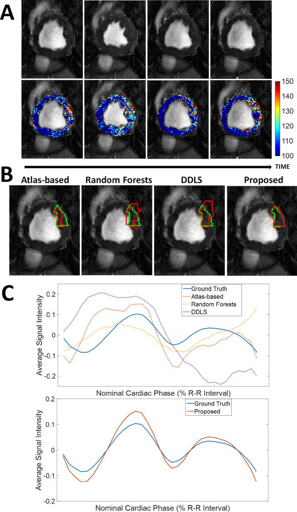
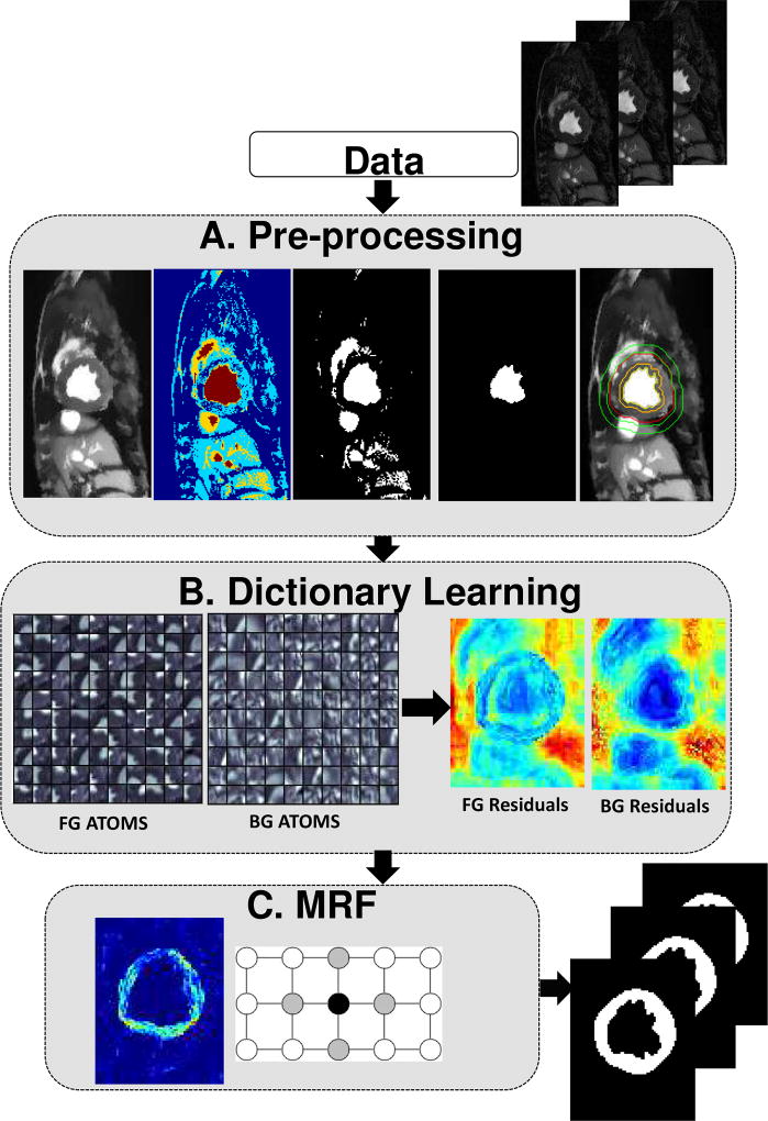
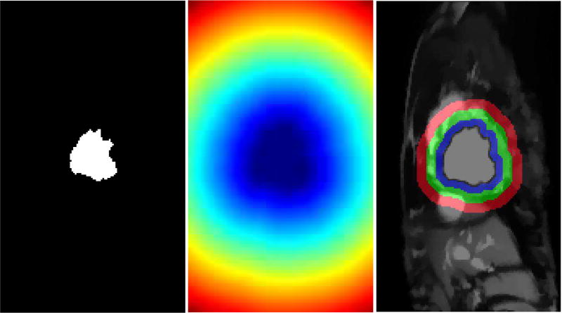
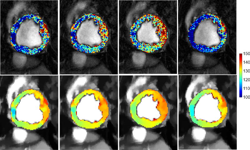
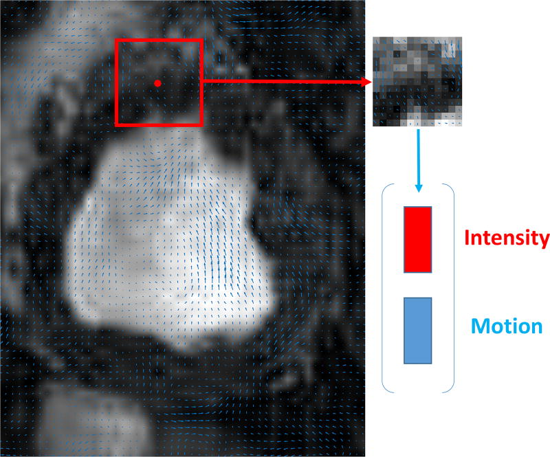
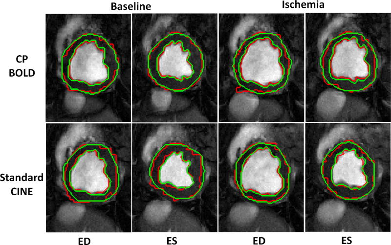
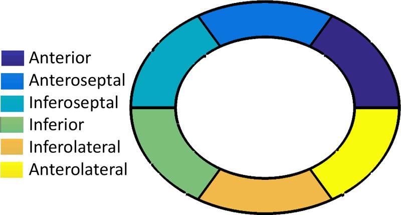
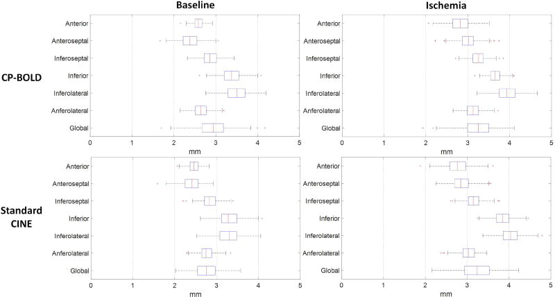
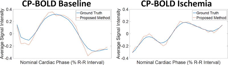
A fully automated 2-D+time myocardial segmentation framework is proposed for cardiac magnetic resonance (CMR) blood-oxygen-level-dependent (BOLD) data sets. Ischemia detection with CINE BOLD CMR relies on spatio-temporal patterns in myocardial intensity, but these patterns also trouble supervised segmentation methods, the de facto standard for myocardial segmentation in cine MRI. Segmentation errors severely undermine the accurate extraction of these patterns. In this paper, we build a joint motion and appearance method that relies on dictionary learning to find a suitable subspace. Our method is based on variational pre-processing and spatial regularization using Markov random fields, to further improve performance. The superiority of the proposed segmentation technique is demonstrated on a data set containing cardiac phase-resolved BOLD MR and standard CINE MR image sequences acquired in baseline and ischemic condition across ten canine subjects. Our unsupervised approach outperforms even supervised state-of-the-art segmentation techniques by at least 10% when using Dice to measure accuracy on BOLD data and performs at par for standard CINE MR. Furthermore, a novel segmental analysis method attuned for BOLD time series is utilized to demonstrate the effectiveness of the proposed method in preserving key BOLD patterns.


Figures

Fig. 1
Fig. 1
BOLD contrast challenges myocardial segmentation algorithms. A: Raw BOLD images from different cardiac phases of the same healthy subject) and color-coded myocardia overlaid on the raw images to demonstrate that subtle, imperceptible to the eye, intensity changes occur. B: Results of various algorithms (shown in red) for myocardial segmentation of the anterior region together with ground truth (green) manual delineations. Algorithms used: Atlas-based [6], Random Forests on Appearance and Texture features (a baseline) and a Dictionary Learning method (DDLS) [7]. C: Corresponding time series of the Anterior region from different methods compared to the one obtained based on ground truth segmentation. Overall errors in segmentation lead to deviations in the estimated time series, which will ultimately lead to low accuracy in ischemia detection. Our proposed method achieves high segmentation accuracy (last image in B); which leads to a better estimate of the time series (bottom part of C). [In typical CP-BOLD acquisition settings, with ECG-triggering, first and last points in the R-R interval correspond to diastole, whereas systole tends to appear around 30%.]
Fig. 2
Fig. 2
Description of the proposed method. Block A aims to find a rough segmentation of the myocardium. In Block B two subject-specific dictionaries are trained on foreground and background on appearance and motion. In Block C a MRF-based segmentation algorithm on the residuals of the two dictionaries is utilized to have smooth boundaries.
Fig. 3
Fig. 3
Extracting candidate background and myocardium regions. LV blood pool (left); Distance transform from the LV blood pool boundary (middle); Rudimentary background and foreground classes (right). Only pixels within the blue and red rings (right panel) are used to sample patches for dictionary learning. The green ring acts as boundary in between these two regions to reduce the chance of false positives.
Fig. 4
Fig. 4
Influence of Total Variation based smoothing on different cardiac phases of a healthy subject. Four temporal phases of the same acquisition of a subject before (top) and after pre-processing (bottom), where myocardial intensities have been color-coded to aid visualization. Observe, how myocardial intensities appear smoother and within the same (and shorter) range across the cardiac cycle after TV-based smoothing (bottom row).
Fig. 5
Fig. 5
The feature vector generation as concatenation of intensities of square patches and corresponding motion vectors inside that patch.
Fig. 6
Fig. 6
Segmentation result (red) of Proposed method for both CP-BOLD MR and standard CINE MR at baseline and ischemic condition for End-diastole (ED) and End-systole (ES) superimposed with corresponding Manual Segmentation (green) contours delineated by experts.
Fig. 7
Fig. 7
Six segments of mid-ventricular myocardial slice
Fig. 8
Fig. 8
Segmental Hausdorff distance accuracy for CP-BOLD and standard CINE MR for epicardium.
Fig. 9
Fig. 9
Normalized time series obtained by averaging pixel intensities in the anterior region, as defined using ground truth (blue) and automatic segmentation (red dotted line) in a subject at baseline (left) and after LAD stenosis and during ischemia (right). Observe that the time series obtained via the proposed segmentation is consistent with that of ground truth, which eventually result in more accurate ischemia detection.
Fig. 10
Fig. 10
Effect of Pre-processing on segmentation accuracy. Rudimentary class thickness is varied from the original size (6mm) for background (a) and myocardium (b). The influence of changing the thickness from 3mm to 9mm of both classes on segmentation accuracy is minimal.
Fig. 11
Fig. 11
Effect of patch size (a), dictionary size (b) and sparsity threshold (c) on segmentation accuracy. The optimal results were obtained using a patch size of 13 × 13, a dictionary of 400 atoms and a sparsity threshold of 4.

References

    1. Dharmakumar R, Arumana JM, Tang R, Harris K, Zhang Z, Li D. Assessment of regional myocardial oxygenation changes in the presence of coronary artery stenosis with balanced SSFP imaging at 3.0T: Theory and experimental evaluation in canines. JMRI. 2008;27(5):1037–1045. - PubMed
    1. Tsaftaris SA, Zhou X, Tang R, Li D, Dharmakumar R. Detecting myocardial ischemia at rest with Cardiac Phase-resolved Blood Oxygen Level-Dependent Cardiovascular Magnetic Resonance. Circ. Cardiovasc. Imaging. 2013;6(2):311–319. - PMC - PubMed
    1. Tsaftaris SA, Tang R, Zhou X, Li D, Dharmakumar R. Ischemic extent as a biomarker for characterizing severity of coronary artery stenosis with blood oxygen-sensitive MRI. JMRI. 2012;35(6):1338–1348. - PMC - PubMed
    1. Bevilacqua M, Dharmakumar R, Tsaftaris SA. Dictionary-driven ischemia detection from cardiac phase-resolved myocardial BOLD MRI at rest. IEEE TMI. 2016;35(1):282–293. - PMC - PubMed
    1. Oksuz I, Mukhopadhyay A, Bevilacqua M, Dharmakumar R, Tsaftaris SA. Dictionary learning based image descriptor for myocardial registration of CP-BOLD MR. MICCAI. 2015:205–213.

Publication types