Review
. 2017 Jul 13;170(2):226-247.
doi: 10.1016/j.cell.2017.06.036.

Evolution of the Human Nervous System Function, Structure, and Development

Affiliations
Review

Evolution of the Human Nervous System Function, Structure, and Development

André M M Sousa et al. Cell. .

Abstract

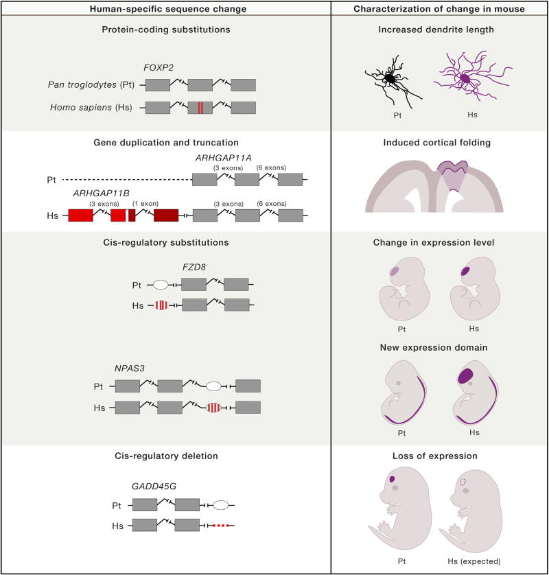
The nervous system-in particular, the brain and its cognitive abilities-is among humans' most distinctive and impressive attributes. How the nervous system has changed in the human lineage and how it differs from that of closely related primates is not well understood. Here, we consider recent comparative analyses of extant species that are uncovering new evidence for evolutionary changes in the size and the number of neurons in the human nervous system, as well as the cellular and molecular reorganization of its neural circuits. We also discuss the developmental mechanisms and underlying genetic and molecular changes that generate these structural and functional differences. As relevant new information and tools materialize at an unprecedented pace, the field is now ripe for systematic and functionally relevant studies of the development and evolution of human nervous system specializations.


Figures

Figure 1
Figure 1. Humans Are Part of the African Great Ape Clade and Have the Largest Brain among Extant Primates
The images depict adult brains of the species indicated at the leaf node. Chimpanzee and bonobo are represented with identical schematics due to their close similarity. The estimated divergence times with respect to humans among great apes (chimpanzee [4.5–7 mya], gorilla [7–10 mya], and orangutan [12–16 mya]), a small ape (gibbon [18–20 mya]), and an Old World monkey (Rhesus macaque [25–33 mya]) are based on unique gap-free sequence identity (adapted from Locke et al. (2011), Yu et al. (2003), and Chen and Li [2001]) and recent analyses of the fossil record (Katoh et al., 2016; Almécija et al., 2013). For simplicity, only one old-world monkey is depicted (Rhesus macaque, the most commonly studied non-human primate); the dashed line represents the approximate split time from the other extant monkey species. ~1 million years ago (mya) implies approximately 1 mya or less.
Figure 2
Figure 2. Variation in the Number of Cerebral Cortical Neurons across Mammals
The shaded region represents the 95% prediction interval for the data points excluding primate and cetacean points, highlighting that the observed number of cortical neurons in primates does not follow the same scaling rules. What scaling rule cetaceans adhere to is an open question that will be resolved as more data become available. Data for this figure were taken from several sources that used different methodologies and data reporting methods. The numbers on cetaceans were compiled from cerebral neocortical samples by Eriksen and Pakkenberg (2007), Walløe et al. (2010), and Mortensen et al. (2014). Information for the chimpanzee data point was obtained from neocortex by Collins et al. (2016). All other data were retrieved from the dataset generated by Herculano-Houzel et al. (2015) from the entire cerebral cortex.
Figure 3
Figure 3. Language-Related Pathways Are Strongly Lateralized and Modified in Humans
(A and B) Language-related pathways connecting Broca’s (B), Geschwind’s (G), and Wernicke’s (W) neocortical territories, reconstructed based on diffusion tensor imaging data. Both hemispheres share the long direct segment and the posterior indirect segments, which connect Broca’s territory in the frontal cortex with Geschwind’s territory in the parietal cortex and connect Geschwind’s territory with Wernicke’s territory in the temporal cortex, respectively. The direct segment is found exclusively in the left hemisphere and connects Broca’s and Wernicke’s territories. (C) Comparison of arcuate fasciculus projections in humans and non-human primates. In humans, projections extend far into the medial and inferior temporal gyri, whereas projections to the chimpanzee temporal lobe are less extensive into the inferior temporal gyrus. In macaques, the projection into the inferior temporal gyrus appears to be completely absent. PFC, prefrontal cortex; SF, Sylvian fissure; STC, superior temporal cortex. Adapted from Catani and Mesulam (2008); Rilling et al. (2008), and Ghazanfar (2008).
Figure 4
Figure 4. Comparative Analysis of Neocortical Gene Expression between Human and Macaque Reveal Species Differences during Fetal Development
(A) Illustration showing gradients of gene co-expression modules (M) during human neocortical development. Four gradients are shown: anterio-posterior (10–13 pcw), ventro-medial (13–16 pcw), temporal (16–19 pcw), and perisylvian (19–24). Genes present within each co-expression module shown here are listed in Pletikos et al. (2014). pcw: postconcepional week; MFC: medial prefrontal cortex; OFC: orbital prefrontal cortex; DFC: dorsolateral prefrontal cortex; VFC: ventrolateral prefrontal cortex; M1C: primary motor cortex; S1C: primary somatosensory cortex; IPC: inferior parietal cortex; STC: superior temporal cortex; ITC: inferior temporal cortex; A1C: primary auditory cortex; V1C: primary visual cortex. (B) Quantitative real-time PCR of hub genes belonging to the coexpression modules represented in (A) show divergence between human and macaque, particularly in the expression of CLMP and C13ORF38. Adapted from Pletikos et al. (2014).
Figure 5
Figure 5. Mouse Models of Human-Specific Genomic Features
Each row presents a study that investigated a different type of human-specific change in a mouse model, with the right column highlighting a specific finding of the study. In order, the studies are Enard et al. (2009), Florio et al. (2015), Boyd et al. (2015), Kamm et al. (2013a), and McLean et al. (2011). The darker shade in ARHGAP11B represents a frameshift and termination that followed a deletion in the fifth exon. Pt, Pan troglodytes, common chimpanzee; Hs, Homo sapiens, human. Note that the findings displayed, particularly for the Enard et al. (2009) and Boyd et al. (2015) studies, are not intended to serve as a summary for all the phenotypes investigated.

Similar articles

Cited by

References

    1. Aiello LC, Wheeler P. The expensive-tissue hypothesis: the brain and the digestive system in human and primate evolution. Curr. Anthropol. 1995;36:199–221.
    1. Allman JM, Tetreault NA, Hakeem AY, Manaye KF, Semendeferi K, Erwin JM, Park S, Goubert V, Hof PR. The von Economo neurons in frontoinsular and anterior cingulate cortex in great apes and humans. Brain Struct. Funct. 2010;214:495–517. - PubMed
    1. Almécija S, Tallman M, Alba DM, Pina M, Moyá-Solá S, Jungers WL. The femur of Orrorin tugenensis exhibits morphometric affinities with both Miocene apes and later hominins. Nat. Commun. 2013;4:2888. - PubMed
    1. Alonso CR, Wilkins AS. The molecular elements that underlie developmental evolution. Nat. Rev. Genet. 2005;6:709–715. - PubMed
    1. Anderson JM, Gilmore R, Roper S, Crosson B, Bauer RM, Nadeau S, Beversdorf DQ, Cibula J, Rogish M, 3rd, Kortencamp S, et al. Conduction aphasia and the arcuate fasciculus: A reexamination of the Wernicke-Geschwind model. Brain Lang. 1999;70:1–12. - PubMed

Substances

LinkOut - more resources