

Animal models of biliary injury and altered bile acid metabolism
- PMID: 28709963
- PMCID: PMC5764833
- DOI: 10.1016/j.bbadis.2017.06.027
Animal models of biliary injury and altered bile acid metabolism
Abstract
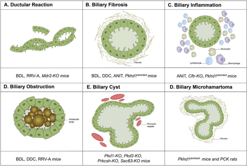
In the last 25years, a number of animal models, mainly rodents, have been generated with the goal to mimic cholestatic liver injuries and, thus, to provide in vivo tools to investigate the mechanisms of biliary repair and, eventually, to test the efficacy of innovative treatments. Despite fundamental limitations applying to these models, such as the distinct immune system and the different metabolism regulating liver homeostasis in rodents when compared to humans, multiple approaches, such as surgery (bile duct ligation), chemical-induced (3,5-diethoxycarbonyl-1,4-dihydrocollidine, DDC, α-naphthylisothiocyanate, ANIT), viral infections (Rhesus rotavirustype A, RRV-A), and genetic manipulation (Mdr2, Cftr, Pkd1, Pkd2, Prkcsh, Sec63, Pkhd1) have been developed. Overall, they have led to a range of liver phenotypes recapitulating the main features of biliary injury and altered bile acid metabolisms, such as ductular reaction, peribiliary inflammation and fibrosis, obstructive cholestasis and biliary dysgenesis. Although with a limited translability to the human setting, these mouse models have provided us with the ability to probe over time the fundamental mechanisms promoting cholestatic disease progression. Moreover, recent studies from genetically engineered mice have unveiled 'core' pathways that make the cholangiocyte a pivotal player in liver repair. In this review, we will highlight the main phenotypic features, the more interesting peculiarities and the different drawbacks of these mouse models. This article is part of a Special Issue entitled: Cholangiocytes in Health and Disease edited by Jesus Banales, Marco Marzioni, Nicholas LaRusso and Peter Jansen.
Keywords: Altered bile acid metabolism; Biliary injury; Cholangiocyte; Experimental models.
Copyright © 2017 Elsevier B.V. All rights reserved.
Figures
References
-
- L. European Association for the Study of the, EASL clinical practice guidelines: management of cholestatic liver diseases. J Hepatol. 2009;51:237–267. - PubMed
-
- Hirschfield GM, Heathcote EJ, Gershwin ME. Pathogenesis of cholestatic liver disease and therapeutic approaches. Gastroenterology. 2010;139:1481–1496. - PubMed
-
- Gossard AA, Talwalkar JA. Cholestatic liver disease. Med Clin North Am. 2014;98:73–85. - PubMed
-
- Pollock G, Minuk GY. Diagnostic considerations for cholestatic liver disease. J Gastroenterol Hepatol. 2017 - PubMed
-
- S B, Fox JG, Davisson MT, et al., editors. The Mouse in Biomedical Research. Elsevier, Place Published; Amsterdam: 2007.
Publication types
MeSH terms
Substances
Grants and funding
LinkOut - more resources
Full Text Sources
Other Literature Sources
Medical
Miscellaneous
