Review
. 2018 Jan 2;8(1):a031211.
doi: 10.1101/cshperspect.a031211.

Regulation of Bone Metabolism by Sex Steroids

Affiliations
Review

Regulation of Bone Metabolism by Sex Steroids

Sundeep Khosla et al. Cold Spring Harb Perspect Med. .

Abstract

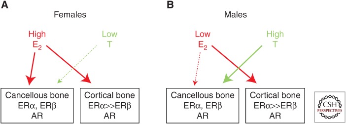
Osteoporosis is a significant public health problem, and a major cause of the disease is estrogen deficiency following menopause in women. In addition, considerable evidence now shows that estrogen is also a major regulator of bone metabolism in men. Since the original description of the effects of estrogen deficiency on bone by Fuller Albright more than 70 years ago, there has been enormous progress in understanding the mechanisms of estrogen and testosterone action on bone using human and mouse models. Although we understand more about the effects of estrogen on bone as compared with testosterone, both sex steroids do play important roles, perhaps in a somewhat compartment-specific (i.e., cancellous vs. cortical bone) manner. This review summarizes our current knowledge of sex steroid action on bone based on human and mouse studies, identifies both agreements and potential discrepancies between these studies, and suggests directions for future research in this important area.


Figures

Figure 1.
Figure 1.
Changes in estradiol, follicle-stimulating hormone (FSH), and bone resorption through the menopausal transition. Population mean bone resorption marker (urine N-telopeptide of type I collagen [NTx]), serum estradiol, and serum FSH levels in relation to years from final menstrual period. Dashed lines denote the 95% confidence intervals. (From Sowers et al. 2013; reproduced, with permission, from Oxford University Press © 2013.)
Figure 2.
Figure 2.
Deconvoluting the effects of estrogen versus testosterone on bone metabolism in men. Percent changes in (A) bone-resorption markers (urinary deoxypyridinoline [Dpd] and N-telopeptide of type I collagen [NTx]), and (B) bone-formation markers (serum osteocalcin and amino-terminal extension peptide of type I collagen [PINP]) in a group of elderly men (mean age 68 yr) made acutely hypogonadal and treated with an aromatase inhibitor (group A), treated with estrogen alone (group B), testosterone alone (group C), or both (group D). See text for details. Asterisks indicate significance for change from baseline: *P < 0.05; **P < 0.01; ***P < 0.001. (From Falahati-Nini et al. 2000; adapted, with permission, from the American Society for Clinical Investigation © 2000.)
Figure 3.
Figure 3.
Effects of interleukin (IL)-1 or tumor necrosis factor (TNF) blockade on bone resorption in women following estrogen withdrawal. Proportional change (%) in serum C-telopeptide of type I collagen (CTx) in postmenopausal women treated for 60 days with transdermal estradiol, made acutely estrogen deficient, and treated with saline (control), an IL-1 blocker (anakinra), or a TNF blocker (etanercept). (From Charatcharoenwitthaya et al. 2007; reproduced, with permission, from John Wiley and Sons © 2007.)
Figure 4.
Figure 4.
Effects of estrogen on cell surface receptor activator of nuclear factor κB ligand (RANKL) expression in postmenopausal women. Changes in osteoprotegerin (OPG)-Fc-FITC fluorescence as an index of mean RANKL surface concentration per cell are shown for premenopausal women (red bars), estrogen-deficient postmenopausal women (green bars), and estrogen-treated postmenopausal women (black bars). (AD) The various cell populations as indicated. P-values by analysis of variance (ANOVA) are as indicated. ***P < 0.001 versus the premenopausal women. (From Eghbali-Fatourechi et al. 2003; reproduced, with permission, from the American Society for Clinical Investigation © 2003.)
Figure 5.
Figure 5.
Working model to explain the differential effects of estrogen versus testosterone on cancellous and cortical bone. Please see text for further details. ER, Estrogen receptor; AR, androgen receptor.

References

    1. Albright F. 1940. Post-menopausal osteoporosis. Trans Assoc Am Physicians 55: 298–305.
    1. Almeida M, Iyer S, Martin-Millan M, Bartell SM, Han L, Ambrogini E, Onal M, Xiong J, Weinstein RS, Jilka RL, et al. 2013. Estrogen receptor-α signaling in osteoblast progenitors stimulates cortical bone accrual. J Clin Invest 123: 394–404. - PMC - PubMed
    1. Almeida M, Laurent MR, Dubois V, Claessens F, O’Brien CA, Bouillon R, Vanderschueren D, Manolagas SC. 2017. Estrogens and androgens in skeletal physiology and pathophysiology. Physiol Rev 97: 135–187. - PMC - PubMed
    1. Bilezikian JP, Morishima A, Bell J, Grumbach MM. 1998. Increased bone mass as a result of estrogen therapy in a man with aromatase deficiency. N Engl J Med 339: 599–603. - PubMed
    1. Bonnick SL. 1998. Skeletal anatomy in densitometry. In Bone densitometry in clinical practice (ed. Bonnick SL), pp. 35–78. Humana, New York.

Publication types

LinkOut - more resources