. 2017 Jul 14;7(1):5379.
doi: 10.1038/s41598-017-05808-w.

RLR-mediated antiviral innate immunity requires oxidative phosphorylation activity

Affiliations

RLR-mediated antiviral innate immunity requires oxidative phosphorylation activity

Takuma Yoshizumi et al. Sci Rep. .

Abstract

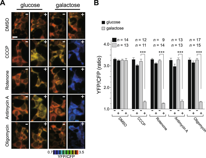
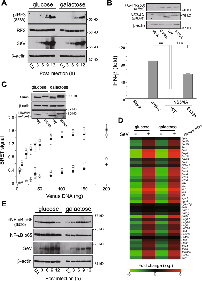
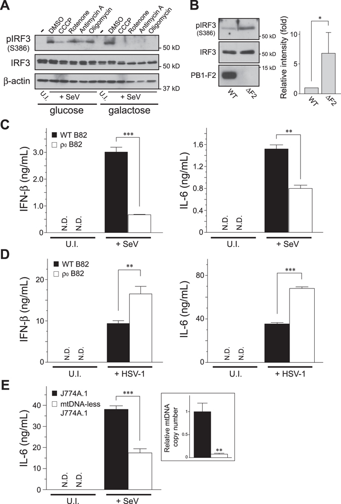
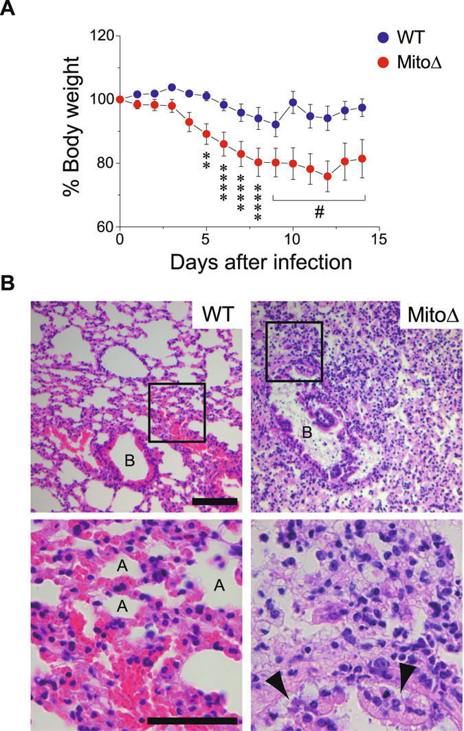
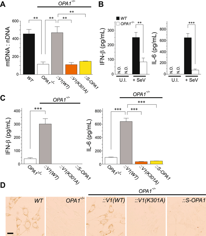
Mitochondria act as a platform for antiviral innate immunity, and the immune system depends on activation of the retinoic acid-inducible gene I (RIG-I)-like receptors (RLR) signaling pathway via an adaptor molecule, mitochondrial antiviral signaling. We report that RLR-mediated antiviral innate immunity requires oxidative phosphorylation (OXPHOS) activity, a prominent physiologic function of mitochondria. Cells lacking mitochondrial DNA or mutant cells with respiratory defects exhibited severely impaired virus-induced induction of interferons and proinflammatory cytokines. Recovery of the OXPHOS activity in these mutants, however, re-established RLR-mediated signal transduction. Using in vivo approaches, we found that mice with OXPHOS defects were highly susceptible to viral infection and exhibited significant lung inflammation. Studies to elucidate the molecular mechanism of OXPHOS-coupled immune activity revealed that optic atrophy 1, a mediator of mitochondrial fusion, contributes to regulate the antiviral immune response. Our findings provide evidence for functional coordination between RLR-mediated antiviral innate immunity and the mitochondrial energy-generating system in mammals.


Conflict of interest statement

The authors declare that they have no competing interests.

Figures

Figure 1
Figure 1
Bioenergetic profiling of cells under oxidative conditions. (A) HEK293 cells stably expressing the ATeam1.03 were cultured in customized media containing either galactose (right) or glucose (left) at 37 °C, and then the indicated mitochondrial inhibitors were added to each medium. The time-course of the fluorescence emission ratio (YFP/CFP) was monitored to visualize cytosolic ATP levels in each living cell, and ratiometric pseudocolor images of cells pre- (−) or post-treated (+) with inhibitors are shown. The images were processed in MetaMorph (Molecular Devices), and the blue color indicates less cytoplasmic ATP. Scale bar, 20 μm. (B) Quantification of the YFP/CFP ratio calculated from images in (A). Number of cells (n) used for the quantification is shown at the top of the graph. Error bars indicate SD (Unpaired t-test; ***P < 0.001).
Figure 2
Figure 2
RLR-mediated signal transduction under oxidative conditions. (A) The kinetic profile of IRF-3 activation in oxidative or glycolytic medium-cultured HEK293 cells that were challenged with SeV (4 HA units/mL). Each cell lysate was collected at the indicated time-points (3, 6, 9 and 12 h) and analyzed by Western blotting with antibodies against the specific antibody (pIRF-3; phosphorylation of Ser386). Anti-β-actin was used as the loading control and anti-SeV as the infection control. U.I., uninfected. (B) Oxidative medium-cultured HEK293 cells were transfected with 50 ng of empty vector (Mock) or expression plasmid for Myc-tagged RIG-I(1–250) (control) together with the IFN-β reporter plasmid. The two right lanes (+NS3/4A) indicate that 100 ng of FLAG-tagged WT or inactive (S139A) NS3/4A serine protease expression plasmids were also co-transfected with the RIG-I(1–250) plasmid. The immunoblot on the top represents an expression profile of Myc-tagged RIG-I(1–250) and FLAG-tagged NS3/4A mutants as well as the loading control of endogenous β-actin. Error bars indicate SD (n = 3; Unpaired t-test; **P < 0.01 and ***P < 0.001, respectively). (C) BRET saturation assay of MAVS oligomerization in glycolytic versus oxidative media. HEK293 cells were co-transfected with 5 ng NLuc-MAVS expression plasmid and increasing amounts (0–200 ng) of Venus-tagged MAVS plasmid along with 200 ng of WT (circles) or S139A (squares) FLAG-tagged NS3/4 A plasmids, and analyzed 24 h later using a BRET saturation assay. Closed and open symbols represent glycolytic and oxidative conditions of HEK293 cells, respectively, and inset blots show Western blots from the BRET saturation point of each curve by immunoblotting with the indicated antibodies. Error bars indicate SEM (n = 3). (D) Heat maps of microarray analysis. Total RNAs were isolated from glycolytic and oxidative cultured conditions of primary MEFs that were unchallenged (−) or challenged (+) with SeV (30 HA units mL−1) for 6 h, and microarray analysis was performed. The heat map was generated by MeV software, and the color indicates the distance from the median of each row. (E) Similar to (A), except that J774A.1 macrophages were challenged with SeV (2 HA units/mL). U.I., uninfected.
Figure 3
Figure 3
OXPHOS activity couples with the RLR pathway to execute antiviral signal transduction. (A) HEK293 cells cultured in glycolytic or oxidative media were infected with SeV (4 HAU/mL) for 2.5 h, and the infected cells were further incubated with the indicated mitochondrial inhibitors for 2.5 h (total 5 h infection). The activation of endogenous IRF-3 was analyzed by Western blotting with antibodies against the specific antibody (pIRF-3; Ser386). Anti-β-actin was used as the loading control. U.I., uninfected. (B) Comparison of the IRF-3 activation between cells infected with recombinant influenza A/PR8 viruses (WT versus ΔF2, each used 2 HAU/mL) cultured under oxidative conditions. The ΔF2 strain is a mutant strain with genetic removal of the PB1-F2 gene from the viral genome. The graph on the right shows the quantification of pIRF-3 bands analyzed by densitometry. Error bars indicate SD (n = 3; Unpaired t-test; *P < 0.05). (C,D) The B82 WT cybrids and ρ0 cells were infected with (C) SeV (4 HAU/mL) for 18 h or (D) HSV-1 (1 × 105 PFU) for 24 h, and the cell-free supernatants were analyzed by ELISA to measure the secreted amounts of IFN-β (left panel) and IL-6 (right panel), respectively. Error bars indicate SD (n = 3; Unpaired t-test; **P < 0.01 and ***P < 0.001, respectively). U.I., uninfected. N.D., not detected. (E) Similar to (C), except that the mtDNA-less J774A.1 and its parental macrophages were infected with SeV (2 HA units/mL). Inset panel: relative mtDNA copy number was confirmed by qPCR. In (CE), cells were maintained in ρ0 medium.
Figure 4
Figure 4
Defects in mtDNA cause malfunction in antiviral innate immunity. (A,B) The 143B cybrids and ρ0 cells were infected with SeV (5 HAU/mL) for 18 h, and (A) activation of both IRF-3 and IκBα was analyzed by Western blotting with antibodies against its specific phosphorylated-detection antibodies (Ser386 for IRF-3 and Ser32/36 for IκBα) or (B) the cell-free supernatants were analyzed by ELISA to measure the secreted amounts of IFN-β (top panel) and IL-6 (bottom panel). U.I., uninfected. Error bars indicate SD (n = 3; Unpaired t-test; ***P < 0.001). (C) Fluorescence microscopy of 143B cybrid cells infected with IAV-GFP (0.6 HAU/mL) for 24 h. In (AC), cells were maintained in ρ0 medium. (D) Similar to (B), except that succinate (suc), the complex II substrate with ADP, was added to the oxidative medium as indicated. Error bars indicate SD (n = 3; Unpaired t-test; ***P < 0.001; N.D., not detected; N.S., not significant).
Figure 5
Figure 5
OXPHOS-defective mice are highly susceptible to viral-infection. (A) Mito-miceΔ (n = 7) or WT mice (n = 9) were challenged with IAV (1 × 103 PFU) and mouse weight loss was monitored for 14 days. In the graph, the percentage change from the initial weight of the mice is shown. Error bars indicate SEM [Dunnett’s test; **P < 0.01 and ****P < 0.0001 (versus day 0), respectively)]. #Dunnett’s test could not be applied because dead mice appeared from day 9. (B) In a separate experiment, the lungs were obtained from each infected mouse on day 8 post-infection, sectioned, and analyzed for histopathology following staining with hematoxylin and eosin. Enlarged boxes are depicted in lower images (400× magnification). Labels A and B in the images indicate alveoli and bronchus, respectively, and arrowheads indicate desquamation of the bronchial epithelium. Scale bars, 100 μm (top) and 50 μm (bottom), respectively.
Figure 6
Figure 6
OPA1 contributes to mitochondrial-mediated antiviral signaling through stabilizing mtDNA. (A) Analysis of mtDNA copy number per nuclear DNA in WT and mutant MEFs. Restoration of mtDNA in OPA1 null cells was measured in mutant cells infected with a retrovirus-expressing variant 1 WT OPA1 [V1(WT)], its K301A mutant [V1(K301A)], and short isoform OPA1 (S-OPA1). Error bars indicate SEM (n = 3; Unpaired t-test; **P < 0.01). (B) SeV-induced antiviral innate immune response in OPA1-null MEFs. The WT and OPA1-null MEFs were either uninfected (U.I.) or infected with SeV (4 HAU/mL) for 18 h, and the cell-free supernatants were analyzed by ELISA to measure the secreted amounts of IFN-β (left panel) and IL-6 (right panel). Error bars indicate SD (n = 3; Unpaired t-test; **P < 0.01 and ***P < 0.001, respectively). N.D., not detected. (C) Similar to (B), except that the immune response in mutant OPA1 MEFs was monitored. Error bars indicate SD (n = 3; Unpaired t-test; ***P < 0.001). (D) Cytochemical analysis of COX activity. Cells expressing COX activity were indicated by a brown color. Scale bar, 20 μm.

References

    1. Hoffmann JA, Kafatos FC, Janeway CA, Jr., Ezekowitz RAB. Phylogenetic perspectives in innate immunity. Science. 1999;284:1313–1318. doi: 10.1126/science.284.5418.1313. - DOI - PubMed
    1. Kawai T, Akira S. Innate immune recognition of viral infection. Nat. Immunol. 2006;7:131–137. doi: 10.1038/ni1303. - DOI - PubMed
    1. Meylan E, Tschopp J. Toll-like receptors and RNA helicases: two parallel ways to trigger antiviral responses. Mol. Cell. 2006;22:561–569. doi: 10.1016/j.molcel.2006.05.012. - DOI - PubMed
    1. Iwasaki A, Medzhitov R. Regulation of adaptive immunity by the innate immune system. Science. 2010;327:291–295. doi: 10.1126/science.1183021. - DOI - PMC - PubMed
    1. Wang C, Youle RJ. The role of mitochondria in apoptosis. Annu. Rev. Genet. 2009;43:95–118. doi: 10.1146/annurev-genet-102108-134850. - DOI - PMC - PubMed

Publication types

MeSH terms