. 2017 Jul 14;7(1):5457.
doi: 10.1038/s41598-017-05657-7.

Plasticity in the Oxidative Folding Pathway of the High Affinity Nerita Versicolor Carboxypeptidase Inhibitor (NvCI)

Affiliations

Plasticity in the Oxidative Folding Pathway of the High Affinity Nerita Versicolor Carboxypeptidase Inhibitor (NvCI)

Sebastián A Esperante et al. Sci Rep. .

Abstract

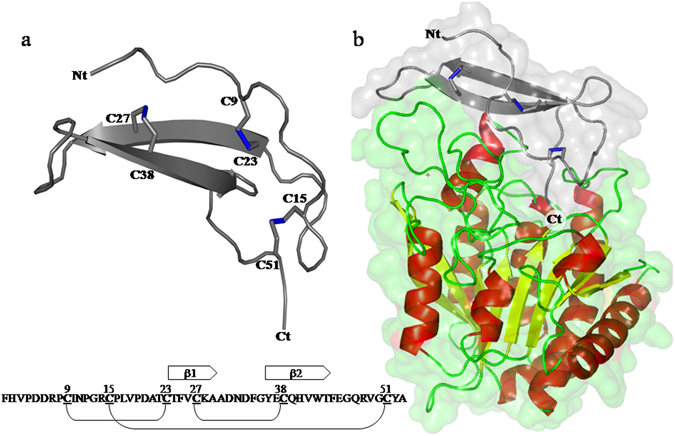
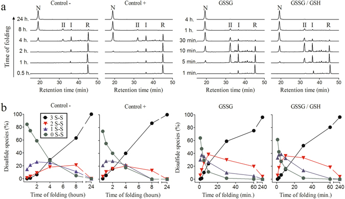
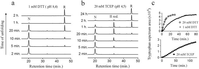
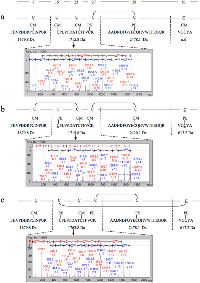
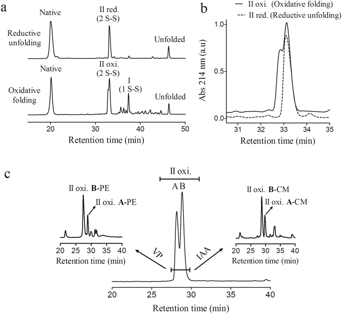
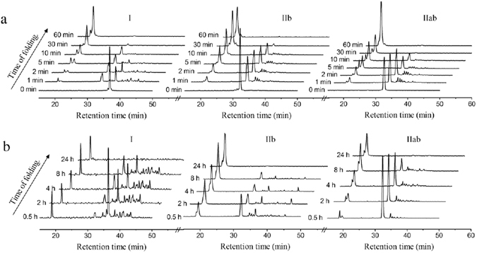
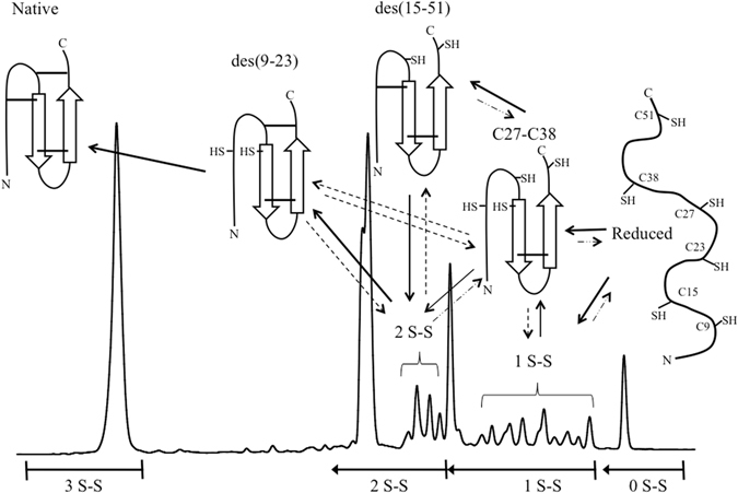
Nerita Versicolor carboxypeptidase inhibitor (NvCI) is the strongest inhibitor reported so far for the M14A subfamily of carboxypeptidases. It comprises 53 residues and a protein fold composed of a two-stranded antiparallel β sheet connected by three loops and stabilized by three disulfide bridges. Here we report the oxidative folding and reductive unfolding pathways of NvCI. Much debate has gone on whether protein conformational folding guides disulfide bond formation or instead they are disulfide bonds that favour the arrangement of local or global structural elements. We show here that for NvCI both possibilities apply. Under physiological conditions, this protein folds trough a funnelled pathway involving a network of kinetically connected native-like intermediates, all sharing the disulfide bond connecting the two β-strands. In contrast, under denaturing conditions, the folding of NvCI is under thermodynamic control and follows a "trial and error" mechanism, in which an initial quasi-stochastic population of intermediates rearrange their disulfide bonds to attain the stable native topology. Despite their striking mechanistic differences, the efficiency of both folding routes is similar. The present study illustrates thus a surprising plasticity in the folding of this extremely stable small disulfide-rich inhibitor and provides the basis for its redesign for biomedical applications.


Conflict of interest statement

The authors declare that they have no competing interests.

Figures

Figure 1
Figure 1
Schematic view of the native three-dimensional structure of NvCI and the NvCI-hCPA4 complex. (a) Schematic view of the ribbon representation of NvCI. The cysteine residues are depicted in the structure and the disulfide bonds are shown in stick representation (blue). The amino acid sequence of NvCI and its secondary structure elements and disulfide pairing are schematically shown at the bottom. The inhibitory site for MCPs comprises two residues (Tyr52 and Ala53), located at the C terminal tail, after Cys51. (b) Surface and ribbon representation of human CPA4 (hCPA4) in complex with NvCI (gray). The three disulfide bridges formed in NvCI are shown in stick representation (blue). The α-helix, β-strands, and coils of hCPA4 are highlighted in red, yellow, and green color, respectively. Ct and Nt stands for C terminus and N terminus, respectively. The Protein Data Bank accession number for the structure of NvCI in complex with hCPA4 is 4A94. All figures were prepared with PyMOL.
Figure 2
Figure 2
RP-HPLC analysis of the folding intermediates and quantitative analysis of disulfide species along the oxidative folding pathway of NvCI. Folding was carried out in Tris-HCl buffer (pH 8.4) in the absence (Control−) and presence of selected redox agents: 0.25 mM 2-mercaptoethanol (Control+), 0.5 mM GSSG or a mixture of 0.5 mM GSSG and 1 mM GSH. (a) Chromatographic profiles of the folding reactions. Intermediates were acid-trapped at the noted times and analyzed by RP-HPLC. Retention times of the native (N) and fully reduced/unfolded (R) forms as well as the two major intermediates (I and II) are indicated. (b) Disulfide species in the oxidative folding of NvCI. Intermediates were trapped by alkylation of the free cysteines at various times and analyzed by MALDI-TOF-MS. 0S-S, 1S-S, 2S-S, and 3S-S represent the completely reduced, one disulfide, two disulfide, and three disulfide species, respectively.
Figure 3
Figure 3
Secondary and tertiary structural changes upon oxidative folding of NvCI. The secondary and tertiary structure changes along folding time were monitored by far-UV CD and tryptophan fluorescence spectra measurements, respectively. The fully reduced/unfolded protein was allowed to refold in 0.1 M Tris-HCl pH 8.4 at 20 °C. (a) Plot of ellipticity at 217 nm as a function of folding time. Inset: Far-UV CD spectra at different time points along folding. (b) Tryptophan spectrum area changes as a function of folding time. Inset: Tryptophan fluorescence spectra at different time points along folding. The arrow indicates the progression of the folding reaction along time.
Figure 4
Figure 4
Reductive unfolding of native NvCI. Native NvCI was treated with: (a) 1 mM DTT in Tris-HCl (pH 8.4) and (b) 20 mM TCEP in sodium acetate (pH 4.5). Time course intermediates were trapped by acidification and analyzed by RP-HPLC. N and R stand for native and reduced NvCI; II for an intermediate that accumulates upon reduction with 20 mM TCEP. (c) Reductive unfolding of native NvCI followed by changes in tryptophan fluorescence spectra, after the addition of reducing agents. Native NvCI was reduced with TCEP (20 mM) in sodium acetate pH 4.5 (graph below) and DTT (1.0 and 20 mM) in Tris.Cl pH 8, 4 (graph above). Tryptophan fluorescence spectra were measured over time and the spectrum area was plotted as a function of time.
Figure 5
Figure 5
Disulfide-pairing determination of the major oxidative folding intermediates of NvCI. CM stands for carboximethyl cysteine (alkylated with IAA) and PE stands for pyridylethyl cysteine (alkylated with VP). The cysteine positions are indicated above the graph. (a) Characterization of fraction I: The purified fraction I was derivatized with IAA, reduced with DTT and further derivatized with VP prior trypsin digestion. The molecular masses determined by MALDI-TOF-MS for the derivatized tryptic peptides are shown. The 1715.8 Da derivatized peptide was sequenced by MALDI-TOF-MS2 and the spectrum characterized by β and γ ion series with the assigned sequence is shown. (b) Fraction IIb characterization: The purified IIab fraction was derivatized with VP and purified by RP-HPLC (see Fig. 5c, II b-PE). Then it was reduced with DTT, derivatized with IAA and subjected to trypsin digestion and MALDI-TOF analysis. The 1715.8 Da fragment was sequenced by MALDI-TOF-MS2 and the spectrum with the assigned sequence is shown. (c) Fraction IIa characterization: The purified IIab fraction was derivatized with IAA and purified by RP-HPLC (see Fig. 5, II a-CM). Then it was reduced with DTT, derivatized with VP and subjected to trypsin digestion and MALDI-TOF analysis. The 1763.8 Da fragment was sequenced by MALDI-TOF-MS2 and the assigned sequence is shown.
Figure 6
Figure 6
Derivatization and HPLC isolation of two 2 S-S folding intermediates of NvCI. (a) Comparison of RP-HPLC profiles of oxidative folding and reductive unfolding. Fraction II of oxidative folding (IIab) and fraction II of reductive unfolding (IIb) elute at the same retention time. The disulfide bond content of all folding intermediates are indicated. (b) Superimposition of oxidative folding and reductive unfolding RP-HPLC chromatograms, showing the elution position and the heterogeneity of fraction IIab. The RP-HPLC was performed using a C4 column as described in methods. (c) The fraction corresponding to IIab, composed at least by two species (a and b), was purified by RP-HPLC in a C8 column, freeze-dried and derivatized either with VP or IAA. Two major species of each derivatized IIab fraction were further purified by RP-HPLC (left and right insets). An inversion of the order of peaks elution was observed after derivatization. PE stands for pyridylethyl species (akylated with VP) and CM stands for carboximethyl species (alkylated with IAA).
Figure 7
Figure 7
Stop/go folding of the major folding/unfolding intermediates of NvCI. Acid-trapped intermediates (I, IIb and IIab) were purified by RP-HPLC, freeze-dried, and dissolved in Tris-HCl (pH 8.4) to reinitiate the folding reaction either in the absence (b) or presence (a) of 1 mM GSSG. Folding intermediates were subsequently trapped with acid at different time points and analyzed by RP-HPLC. The chromatograms at 0 minutes (shown in a) were obtained after resuspending the lyophilized intermediates in TFA 1% as a control of purity and homogeneity of each species.
Figure 8
Figure 8
Oxidative folding of NvCI in the presence of high denaturing conditions. The fully reduced/unfolded protein was allowed to refold in Tris-HCl buffer (pH 8.4) in the presence of: 6.0 M urea; 1 mM GSSG in 6.0 M urea; 4.0 M Gdn.HCl and 1 mM GSSG in 4.0 M Gdn.HCl. Intermediates were acid-trapped at the noted times and analyzed by RP-HPLC. The elution positions of native (N) and reduced (R) NvCI are indicated.
Figure 9
Figure 9
Disulfide scrambling of NvCI under different concentrations of denaturants. (a) The native form of NvCI was denatured in Tris-HCl buffer (pH 8.4) containing 0.25 mM 2-mercaptoethanol as thiol initiator and the indicated concentration of denaturants at 20 °C for 20 h. The denatured samples were quenched with 2% TFA and analyzed by RP-HPLC. The fractions a, b and c correspond to non-native (scrambled) isomers of NvCI. (b) Native fraction as a function of denaturant concentration. Denaturation of native NvCI is defined by the conversion of the native structure into scrambled isomers. The denaturants are Gdn.SCN (⚫), Gdn.HCl (○) and urea (▲).
Figure 10
Figure 10
Schematic representation of the disulfide oxidative folding pathway of NvCI. The polypeptide backbone and the β-sheets of the protein are depicted by lines and arrows, respectively. The positions of the six cysteine residues are indicated (SH: 9, 15, 23, 27, 38 and 51) and the disulfide bonds are depicted as solid lines. The RP-HPLC chromatogram and the major disulfide intermediates are depicted and the disulfide bond content of all the RP-HPLC fractions are indicated below the graph. The solid lines represent the productive disulfide folding pathway proposed.

Similar articles

Cited by

References

    1. Dill KA, Chan HS. From Levinthal to pathways to funnels. Nature structural biology. 1997;4:10–19. doi: 10.1038/nsb0197-10. - DOI - PubMed
    1. Bryngelson JD, Onuchic JN, Socci ND, Wolynes PG. Funnels, pathways, and the energy landscape of protein folding: a synthesis. Proteins. 1995;21:167–195. doi: 10.1002/prot.340210302. - DOI - PubMed
    1. Creighton TE. The protein folding problem. Science. 1988;240(267):344. - PubMed
    1. Gruebele M. The fast protein folding problem. Annu. Rev. Phys. Chem. 1999;50:485–516. doi: 10.1146/annurev.physchem.50.1.485. - DOI - PubMed
    1. Fetrow JS, Giammona A, Kolinski A, Skolnick J. The protein folding problem: a biophysical enigma. Curr. Pharm. Biotechnol. 2002;3:329–347. doi: 10.2174/1389201023378120. - DOI - PubMed

Publication types

MeSH terms

LinkOut - more resources