. 2017 Jul 14;7(1):5491.
doi: 10.1038/s41598-017-05618-0.

Persistent circulation of Coxsackievirus A6 of genotype D3 in mainland of China between 2008 and 2015

Affiliations

Persistent circulation of Coxsackievirus A6 of genotype D3 in mainland of China between 2008 and 2015

Yang Song et al. Sci Rep. .

Abstract

A total of 807 entire VP1 sequences of Coxsackievirus A6 (CV-A6) from mainland of China from 1992 to 2015, including 520 in this study and 287 from the GenBank database, were analysed to provide a basic framework of molecular epidemiological characteristics of CV-A6 in China. Sixty-five VP1 sequences including 46 representative CV-A6 isolates from 807 Chinese strains and 19 international strains from GenBank were used for describing the genotypes and sub-genotypes. The results revealed that CV-A6 strains can be categorised into 4 genotypes designated as A, B, C, and D according to previous data and can be further subdivided into B1-B2, C1-C2, and D1-D3 sub-genotypes. D3 is the predominant sub-genotype that circulated in recent years in mainland of China and represents 734 of 807 Chinese isolates. Sixty-six strains belong to D2, whereas B1 and C1 comprise a single strain each, and five AFP strains formed B2. Sub-genotype D3 first circulated in 2008 and has become the predominant sub-genotype since 2009 and then reached a peak in 2013, while D2 was mostly undetectable in the past years. These data revealed different transmission stages of CV-A6 in mainland of China and that sub-genotype D3 may have stronger transmission ability.


Conflict of interest statement

The authors declare that they have no competing interests.

Figures

Figure 1
Figure 1
Geographic and annual distribution of 807 Chinese CV-A6 strains included in this study.
Figure 2
Figure 2
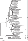
Phylogenetic trees based on the complete VP1 nucleotide sequences of CV-A6 of 65 representative strains isolated between 1949 (the prototype Gdula strain) and 2015. Numbers at the nodes indicate bootstrap support for each node (percentage of 1000 bootstrap replicates). A difference of at least 15% in the entire VP1 region of CV-A6 strains was used to distinguish genotypes. CV-A6 strains are labelled using the following format: ‘isolate name’/‘country of origin’/‘year of isolation’ (details are listed in Table 1). Different genotypes and sub-genotypes are shown to the right of the tree and the scale at the bottom indicates measurement of relative phylogenetic distance. Abbreviations of countries/Province: CHN, China; JPN, Japan; IND, India; TW, Taiwan, China; FIN, Finland; FRA, France; ESP, Spain.
Figure 3
Figure 3
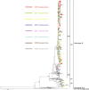
Phylogenetic analyses based on 807 entire VP1 sequences of CV-A6 in China. The phylogenetic tree shows the evolutionary tendency of sub-genotype D2 and D3 strains in China since 2008; sub-genotype D3 was the predominant one. The branches of sequences from mainland of China are highlighted in different colours according to years from 2008 to 2015, the other international strains (including Taiwan strains) and strains detected before 2008 are uncoloured. The phylogenetic tree indicates that evolutionary branch D3a was responsible for the recent outbreaks since 2013. A ladder-like topology revealed that adaptation of CV-A6 occurred during the epidemic history. A prototype CV-A 2 strain (Fleetwood) served as an outgroup.
Figure 4
Figure 4
Yearly proportional distribution of HFMD-associated sub-genotypes D2 and D3 (evolutionary branches D3a and D3b) from 2008 to 2015 in mainland of China.
Figure 5
Figure 5
The geographic distribution of HFMD-associated CV-A6 strains in 7 representative regions of China, the region and the number of the sequences are coloured according to the legend (Taiwan sequences are not included). The free map source was acquired from d-maps.com (URL: http://d-maps.com/carte.php?num_car=15272&lang=en; the content is properly attributed in the appropriate figure legend according to the attribution guidelines; the website is protected by Copyright France, Registration Number: 58KU297). The map was then generated and modified using Microsoft Excel 2010 software.

References

    1. Picornaviridae. Virus Taxonomy: Classification and Nomenclature of Viruses: Ninth Report of the International Committee on Taxonomy of Viruses. (Elsevier, San Diego, 2011).
    1. Oberste MS, Penaranda S, Maher K, Pallansch MA. Complete genome sequences of all members of the species Human enterovirus A. J Gen Virol. 2004;85:1597–1607. doi: 10.1099/vir.0.79789-0. - DOI - PubMed
    1. Oberste MS, et al. Enteroviruses 76, 89, 90 and 91 represent a novel group within the species Human enterovirus A. J Gen Virol. 2005;86:445–451. doi: 10.1099/vir.0.80475-0. - DOI - PubMed
    1. Ayukekbong J, et al. Shift of Enterovirus species among children in Cameroon - Identification of a new enterovirus, EV-A119. J Clin Virol. 2013;58:227–232. doi: 10.1016/j.jcv.2013.07.005. - DOI - PubMed
    1. Razafindratsimandresy, R., Joffret, M. L., Delpeyroux, F. & Heraud, J. M. First full genome sequence of a human enterovirus a120, isolated in madagascar. Genome announcements2, doi:10.1128/genomeA.00568-14 (2014). - PMC - PubMed

Publication types

LinkOut - more resources