. 2017 Jul 14;7(1):5377.
doi: 10.1038/s41598-017-05760-9.

The LRRK2 G2385R variant is a partial loss-of-function mutation that affects synaptic vesicle trafficking through altered protein interactions

Affiliations

The LRRK2 G2385R variant is a partial loss-of-function mutation that affects synaptic vesicle trafficking through altered protein interactions

Maria Dolores Perez Carrion et al. Sci Rep. .

Abstract

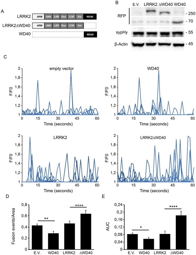
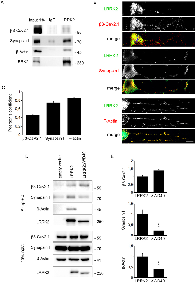
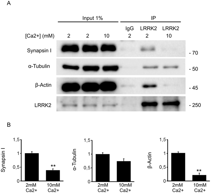
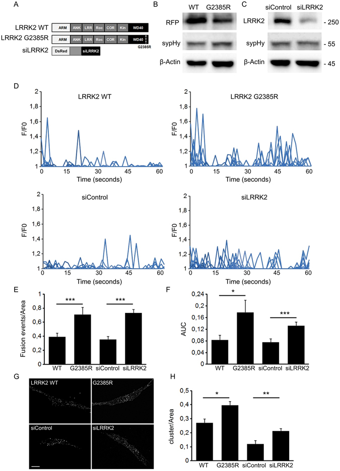
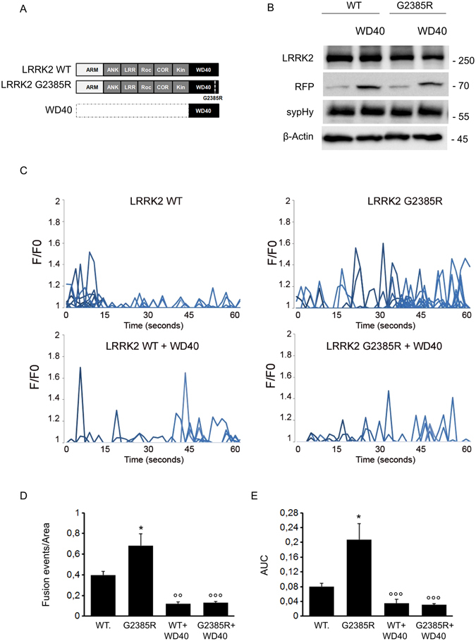
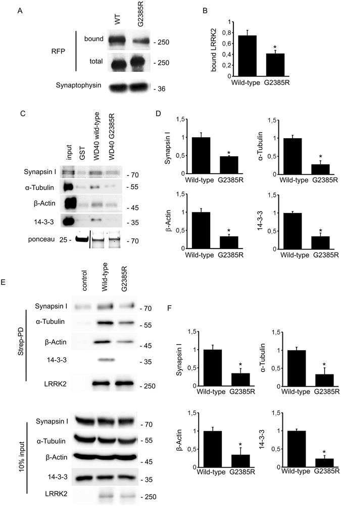
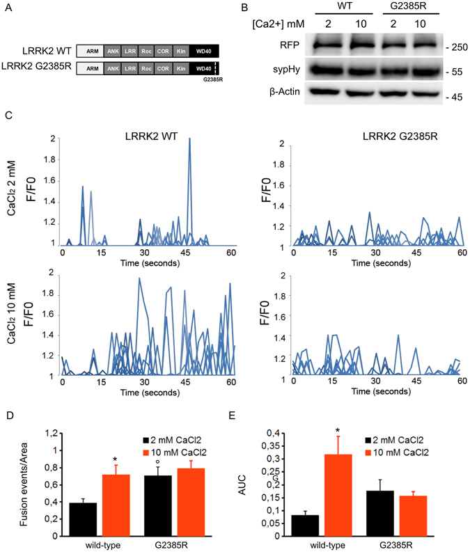
Mutations in the Leucine-rich repeat kinase 2 gene (LRRK2) are associated with familial Parkinson's disease (PD). LRRK2 protein contains several functional domains, including protein-protein interaction domains at its N- and C-termini. In this study, we analyzed the functional features attributed to LRRK2 by its N- and C-terminal domains. We combined TIRF microscopy and synaptopHluorin assay to visualize synaptic vesicle trafficking. We found that N- and C-terminal domains have opposite impact on synaptic vesicle dynamics. Biochemical analysis demonstrated that different proteins are bound at the two extremities, namely β3-Cav2.1 at N-terminus part and β-Actin and Synapsin I at C-terminus domain. A sequence variant (G2385R) harboured within the C-terminal WD40 domain increases the risk for PD. Complementary biochemical and imaging approaches revealed that the G2385R variant alters strength and quality of LRRK2 interactions and increases fusion of synaptic vesicles. Our data suggest that the G2385R variant behaves like a loss-of-function mutation that mimics activity-driven events. Impaired scaffolding capabilities of mutant LRRK2 resulting in perturbed vesicular trafficking may arise as a common pathophysiological denominator through which different LRRK2 pathological mutations cause disease.


Conflict of interest statement

The authors declare that they have no competing interests.

Figures

Figure 1
Figure 1
Domain-wise dissection of LRRK2 impact on vesicle trafficking. (A) Schematic representation of RFP-LRRK2 derived constructs. The distinct LRRK2 domains are indicated. Protein-protein domains: ARM, armadillo repeats; ANK, ankyrin repeats; LRRs, leucine-rich repeats, WD40, WD40 repeats; Roc, Ras of complex proteins; COR, C-terminal of ROC, Kin: kinase domain. (B) Western-blotting analysis of cells expressing synaptopHluorin reporter (sypHy) together with RFP-LRRK2 derived constructs or empty vector (E.V.). (C) Time course analysis of fusion events occurring in SH-5YSY cells transfected with the different LRRK2 derived constructs. SH-5YSY cells were co-transfected with sypHy reporter and empty vector (E.V.) or the indicated RFP-LRRK2 derived constructs. TIRFM imaging was performed 48 hours after transfection. Peaks of fluorescence intensity correspond to single fusion events. Fluorescence data are expressed as F/F0. The graphs show the total number of fusion events (D) and the resulting fluorescence changes (E) expressed as Area Under Curve (AUC) for each construct. Data are normalized for the cell area and are expressed as mean ± SE; n = 20 cells per construct. *p < 0.05, **p < 0.01, ****p < 0.001 ANOVA.
Figure 2
Figure 2
LRRK2 interacts with proteins involved in vesicle trafficking. (A) Immunoprecipitation of endogenous LRRK2 from adult forebrain mouse lysate shows that LRRK2 interacts with β3-Cav2.1, synapsin I and β-Actin. (B) DIV14 cortical neurons were processed for imaging purposes and stained with specific antibodies recognizing LRRK2, β3-Cav2.1, Synapsin I and F-Actin. (C) Graph shows the Pearson’s correlation coefficient to appreciate the overlap between LRRK2 and the indicated protein. (D) We isolated on streptavidin resin full-length strep-FLAG-LRRK2 and strep-FLAG-LRRK2ΔWD40 protein from N2A over-expressing cells. Interacting proteins were resolved by western-blotting. (E) We evaluated the extent of β3-Cav2.1, Synapsin I and β-Actin bound to the different LRRK2 variants. Data are expressed as optical density and normalized versus amount of precipitated LRRK2 protein. Graphs report mean ± S.E, *p < 0.05, Student’s T-test, n = 4.
Figure 3
Figure 3
LRRK2 interactome is shaped by neuronal activity. (A) Immunoprecipitation of endogenous LRRK2 from DIV14 cortical culture incubated with 2 or 10 mM CaCl2 for 30 minutes. (B) We evaluated the extent of synapsin I, α-tubulin and β-actin bound to LRRK2 at 2 and 10 mM CaCl2. Data are expressed as optical density and normalized versus amount of precipitated LRRK2 protein. Graphs report mean ± S.E, **p < 0.01, Student’s T-test, n = 4.
Figure 4
Figure 4
G2385R mutation affects vesicle trafficking. (A) Schematic representation of LRRK2 wild-type and G2385R variant. The distinct LRRK2 domains are indicated. (B) Western-blotting analysis of cells expressing sypHy reporter together with RFP-LRRK2 derived constructs. (C) Western-blotting analysis of cells expressing sypHy reporter together with vector coding for DsRed fluorescent protein and shRNA targeting hLRRK2 (siLRRK2) or a control sequence (siControl). (D) Time course analysis of synaptic events occurring in SH-5YSY cells transfected with wild-type LRRK2 (WT), G2385R-LRRK2, siControl or siLRRK2 under resting conditions. SH-5YSY cells were co-transfected with sypHy and the indicated constructs and imaged by TIRFM 48 hours later. Peaks of variable fluorescence intensity correspond to single fusion events. Fluorescence data are expressed as F/F0. The graphs show the total number of fusion events (E) and the resulting fluorescence changes (F) expressed as Area Under Curve (AUC) for each construct. Data are normalized for the cell area and are the mean ± SE of up to 20 cells per construct. *p < 0.05, ***p < 0.001 ANOVA. (G) Vesicle density in the TIRFM zone after NH4Cl2 treatment. The cells were incubated with the membrane permeant NH4Cl2 solution for 5 minutes, to label all sypHy positive cluster. Then, cells were fixed and imaged by TIRFM to visualize vesicles docked to the plasma membrane. Each spot corresponds to a sypHy positive clusters. Scale bar = 10 μm. (H) The graph reports the number of sypHy positive clusters present under the TIRF zone. Data are normalized for the cell area and are expressed as mean ± SE; n = 15 cells for construct. *p < 0.05, **p < 0.01 ANOVA.
Figure 5
Figure 5
WD40 expression rescues G2385R impact on vesicle trafficking. (A) Schematic representation of LRRK2 wild-type and G2385R variant and isolated WD40 domain. The distinct LRRK2 domains are indicated. (B) Western-blotting analysis of cells expressing sypHy reporter together with RFP-LRRK2 derived constructs. (C) Time course analysis of fusion events occurring in stable N2A line expressing wild-type LRRK2 (WT) or G2385R-LRRK2 and transfected with sypHy together with empty vector or RFP-WD40. N2A cells were co-transfected with sypHy and the indicated constructs and imaged by TIRFM 48 hours later. Peaks of variable fluorescence intensity correspond to single fusion events. Fluorescence data are expressed as F/F0. The graphs show the total number of fusion events (D) and the resulting fluorescence changes (E) expressed as Area Under Curve (AUC) for each construct. Data are normalized for the cell area and are the mean ± SE of up to 20 cells per construct. *p < 0.05 versus LRRK2 wild-type; °°p < 0.01, °°°p < 0.001 WD40 versus LRRK2 WT or G2385R expressing cell, ANOVA.
Figure 6
Figure 6
G2385R mutation affects LRRK2 binding properties. (A) We measured the extent of LRRK2 and SV binding by ultracentrifugation sedimentation assay. We incubated purified RFP-LRRK2 wild-type and G2385R variant with isolated SV (10 μg protein/sample). Bound RFP-LRRK2 was separated from free RFP-LRRK2 by high-speed centrifugation. We appreciated SV-bound LRRK2 by immunoblotting with anti-RFP antibody. The recovery of SV in the pellet was evaluated based on synaptophysin immunoreactivity. (B) The binding of RFP-LRRK2 wild-type and G2385R to SV was calculated as the ratio of total RFP-LRRK2 and expressed as mean ± SE; n = 6. *p < 0.05; Student’s t-test. (C) GST-pull down approach was performed to explore the interactome associated to LRRK2 WD40 domain. (Upper panels) GST-fusion proteins corresponding to WD40 domain of LRRK2 wild type and LRRK2 G2385R were used to retain interactors from adult forebrain lysate. (Lower panel) Ponceau staining shows the abundance of GST fusion proteins used as bait. (D) We evaluated the extent of synapsin I, α-tubulin, β-Actin and 14-3-3 bound to WD40 G2385R domain. Data are expressed as ratio over WD40 wild-type domain. Graph reports mean ± S.E; n = 4. *p < 0.05, Student’s T-test. (E) We isolated on streptavidin resin full-length FLAG-LRRK2 wild-type and FLAG-LRRK2 G2385R protein from N2A over-expressing cells. Interacting proteins were resolved by western-blotting. (F) We evaluated the extent of synapsin I, α-tubulin, β-Actin and 14-3-3 bound to the different LRRK2 variant. Data are expressed as the ratio over LRRK2 wild-type. Graphs report mean ± S.E; n = 4. *p < 0.05, Student’s T-test.
Figure 7
Figure 7
The G2385R variant mimics activity-driven events. (A) Schematic representation of LRRK2 wild-type and G2385R variant. The distinct LRRK2 domains are indicated. (B) Western-blotting analysis of cells expressing sypHy reporter together with RFP-LRRK2 derived constructs upon incubation with 2 or 10 mM CaCl2. (C) Time course analysis of synaptic events occurring in SH-5YSY cells cells transfected with wild-type LRRK2 (WT) or G2385R-LRRK2 upon incubation with 2 or 10 mM CaCl2 (20 minutes in KRH solution). SH-5YSY cells were co-transfected with sypHy and the indicated constructs and imaged by TIRFM 48 hours later. Peaks of variable fluorescence intensity correspond to single fusion events. Fluorescence data are expressed as F/F0. The graphs show the total number of fusion events (D) and the resulting fluorescence changes (E) expressed as Area Under Curve (AUC) for each construct. Data are normalized for the cell area and are the mean ± SE of up to 20 cells per construct. *p < 0.05 10 mM versus 2 mM CaCl2, °p < 0.05 G2385R versus LRRK2 wild-type, 2 mM CaCl2, ANOVA.

References

    1. Wakabayashi K, et al. The Lewy body in Parkinson’s disease and related neurodegenerative disorders. Mol. Neurobiol. 2013;47:495–508. doi: 10.1007/s12035-012-8280-y. - DOI - PubMed
    1. Schulz JB, Hausmann L, Hardy J. 199 years of Parkinson disease - what have we learned and what is the path to the future? J. Neurochem. 2016 - PubMed
    1. Hernandez DG, Reed X, Singleton AB. Genetics in Parkinson disease: Mendelian versus non-Mendelian inheritance. J. Neurochem. 2016 - PMC - PubMed
    1. Civiero L, Dihanich S, Lewis PA, Greggio E. Genetic, structural, and molecular insights into the function of ras of complex proteins domains. Chem. Biol. 2014;21:809–818. doi: 10.1016/j.chembiol.2014.05.010. - DOI - PMC - PubMed
    1. Cirnaru MD, et al. LRRK2 kinase activity regulates synaptic vesicle trafficking and neurotransmitter release through modulation of LRRK2 macro-molecular complex. Front Mol Neurosci. 2014;7:49. doi: 10.3389/fnmol.2014.00049. - DOI - PMC - PubMed

Publication types

MeSH terms