. 2018 Jan;27(1):14-25.
doi: 10.1002/pro.3235. Epub 2017 Sep 6.

UCSF ChimeraX: Meeting modern challenges in visualization and analysis

Affiliations

UCSF ChimeraX: Meeting modern challenges in visualization and analysis

Thomas D Goddard et al. Protein Sci. 2018 Jan.

Abstract

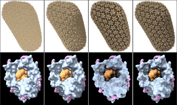
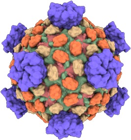
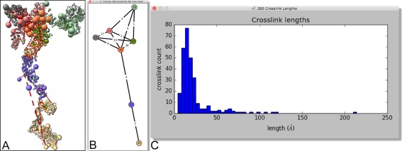
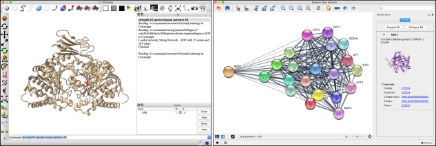
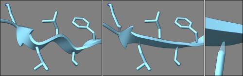
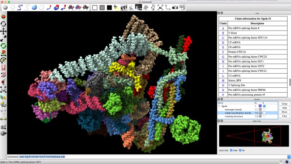
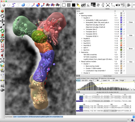
UCSF ChimeraX is next-generation software for the visualization and analysis of molecular structures, density maps, 3D microscopy, and associated data. It addresses challenges in the size, scope, and disparate types of data attendant with cutting-edge experimental methods, while providing advanced options for high-quality rendering (interactive ambient occlusion, reliable molecular surface calculations, etc.) and professional approaches to software design and distribution. This article highlights some specific advances in the areas of visualization and usability, performance, and extensibility. ChimeraX is free for noncommercial use and is available from http://www.rbvi.ucsf.edu/chimerax/ for Windows, Mac, and Linux.

Keywords: biomolecular visualization and analysis; density maps; integrative hybrid modeling; interactive molecular graphics; large-scale data; light-sheet microscopy; structural biology; virtual reality.


Figures

Figure 1
Figure 1
ChimeraX lighting modes. HIV‐1 capsid (PDBid: 3j3q) as all atoms (top row) and cyclodextrin‐admitting outer membrane protein (PDBid: 4d5d) chain A as molecular surfaces (bottom row) in different lighting modes, from left to right: (1) simple lighting, with key, fill, and ambient lights, but no shadows; (2) simple lighting plus directional shadows; (3) ambient occlusion (ambient‐only light, ambient shadowing from 64 directions); (4) full lighting, with key, fill, and ambient lights, directional shadows, and ambient shadowing.
Figure 2
Figure 2
Silhouettes and flat lighting. Nucleosome (PDBid: 5jrg) with DNA as molecular surfaces and histone proteins as helix‐tube cartoons, left: simple lighting with silhouettes; right: flat (ambient‐only) lighting with silhouettes.
Figure 3
Figure 3
Low‐resolution surfaces. Reovirus capsid proteins (PDBid: 1ej6) shown with 15‐Å‐resolution Gaussian surfaces.
Figure 4
Figure 4
Contact plot. (A) CRISPR molecular assembly, two copies in the asymmetric unit of X‐ray structure (PDBid: 5cd4). (B) Schematic showing contacts between chains. (C) Interface between RNA (blue) and one CasC protein shown as solvent‐excluded surface (white) with colored contact residues. (D) Residue schematic of RNA/CasC interface.
Figure 5
Figure 5
Chemical crosslink plots. (A) Integrative hybrid model of nuclear pore subcomplex nup84 with crosslinks shown as dashed lines. (B) Schematic showing numbers of inter‐ and intra‐protein crosslinks. (C) Histogram of crosslink lengths.
Figure 6
Figure 6
Results of executing the ChimeraX stringdb tool to find protein–protein interaction partners for IDH1 (PDBid: 1t09) and display them in Cytoscape.
Figure 7
Figure 7
Ribbon and side chains of a β‐strand in chemoreceptor protein (PDBid: 2gbp), from left to right: unsmoothed, smoothed with tethers, tether closeup.
Figure 8
Figure 8
Typical ChimeraX window, with structure (PDBid: 5gmk) in the main 3D display. Toolbars lie across the top and along the left, command and status lines across the bottom. Along the right are three docked tool windows, from top to bottom: Log, Models, and Side View. The Log is showing an HTML table containing chain information, where the chain letters are clickable links that will bring up the sequences of those chains. The Side View contains its own 3D depiction of the structure from the side.
Figure 9
Figure 9
Virtual‐reality view of molecular structure. Dual with hand controllers (not shown) are used to rotate and scale the image or to position a pointer (yellow cone) to select icons for controlling display styles and colors.
Figure 10
Figure 10
Integrated modeling. ChimeraX window showing starting models, restraints and output from integrative hybrid modeling of the nup84 nuclear pore subcomplex. Hierarchical data organization is shown in upper right including starting comparative models, template structures and sequence alignments for seven components, chemical crosslinking restraints and 2D electron microscopy, and coarse‐grained output ensembles. Graphics shows a best‐scoring model satisfying restraints, large spheres for multi‐residue unordered segments, dashed lines for crosslinks, 2D electron microscopy in background, localization density envelopes for the ensemble of solutions. Lower right panels control electron microscopy rendering and show comparative model and template sequence alignments.

References

    1. O'Donoghue SI, Goodsell DS, Frangakis AS, Jossinet F, Laskowski RA, Nilges M, Saibil HR, Schafferhans A, Wade RC, Westhof E, Olson AJ (2010) Visualization of macromolecular structures. Nat Methods 7:42. - PMC - PubMed
    1. Wang J, Moore PB (2017) On the interpretation of electron microscopic maps of biological macromolecules. Protein Sci 26:122–129. - PMC - PubMed
    1. Baker ML, Zhang J, Ludtke SJ, Chiu W (2010) Cryo‐EM of macromolecular assemblies at near‐atomic resolution. Nat Protoc 5:1697–1708. - PMC - PubMed
    1. Baker ML, Hryc CF, Zhang Q, Wu W, Jakana J, Haase‐Pettingell C, Afonine PV, Adams PD, King JA, Jiang W, Chiu W (2013) Validated near‐atomic resolution structure of bacteriophage epsilon15 derived from cryo‐EM and modeling. Proc Natl Acad Sci USA 110:12301–12306. - PMC - PubMed
    1. Chen BC, Legant WR, Wang K, Shao L, Milkie DE, Davidson MW, Janetopoulos C, Wu XS, Hammer JA, 3rd , Liu Z, English BP, Mimori‐Kiyosue Y, Romero DP, Ritter AT, Lippincott‐Schwartz J, Fritz‐Laylin L, Mullins RD, Mitchell DM, Bembenek JN, Reymann AC, Bohme R, Grill SW, Wang JT, Seydoux G, Tulu US, Kiehart DP, Betzig E (2014) Lattice light‐sheet microscopy: imaging molecules to embryos at high spatiotemporal resolution. Science 346:1257998. - PMC - PubMed

Publication types