. 2017 Nov;154(5):1601-1610.e3.
doi: 10.1016/j.jtcvs.2017.06.035. Epub 2017 Jun 21.

Cardiac reprogramming factor Gata4 reduces postinfarct cardiac fibrosis through direct repression of the profibrotic mediator snail

Affiliations

Cardiac reprogramming factor Gata4 reduces postinfarct cardiac fibrosis through direct repression of the profibrotic mediator snail

Megumi Mathison et al. J Thorac Cardiovasc Surg. 2017 Nov.

Abstract

Objective: The administration of a variety of reprogramming factor cocktails has now been shown to reprogram cardiac fibroblasts into induced cardiomyocyte-like cells. However, reductions in ventricular fibrosis observed after reprogramming factor administration seem to far exceed the extent of induced cardiomyocyte-like cell generation in vivo. We investigated whether reprogramming factor administration might primarily play a role in activating antifibrotic molecular pathways.

Methods: Adult rat cardiac fibroblasts were infected with lentivirus encoding the transcription factors Gata4, Mef2c, or Tbx5, all 3 vectors, or a green fluorescent protein control vector. Gene and protein expression assays were performed to identify relevant antifibrotic targets of these factors. The antifibrotic effects of these factors were then investigated in a rat coronary ligation model.

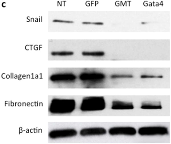
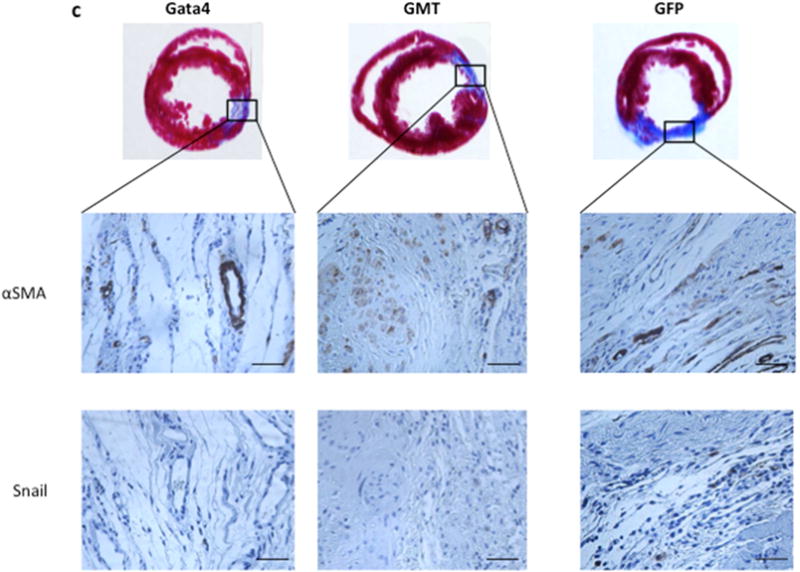
Results: Gata4, Mef2c, or Tbx5 administration to rat cardiac fibroblasts in vitro significantly downregulated expression of Snail and the profibrotic factors connective tissue growth factor, collagen1a1, and fibronectin. Of these factors, Gata4 was shown to be the one responsible for the downregulation of the profibrotic factors and Snail (mRNA expression fold change relative to green fluorescent protein for Snail, Gata4: 0.5 ± 0.3, Mef2c: 1.3 ± 1.0, Tbx5: 0.9 ± 0.5, Gata4, Mef2c, or Tbx5: 0.6 ± 0.2, P < .05). Chromatin immunoprecipitation quantitative polymerase chain reaction identified Gata4 binding sites in the Snail promoter. In a rat coronary ligation model, only Gata4 administration alone improved postinfarct ventricular function and reduced the extent of postinfarct fibrosis.

Conclusions: Gata4 administration reduces postinfarct ventricular fibrosis and improves ventricular function in a rat coronary ligation model, potentially as a result of Gata4-mediated downregulation of the profibrotic mediator Snail.

Keywords: Gata4; Snail; cardiac reprogramming; fibrosis.


Figures

Figure 1
Figure 1
Expression of Snail in cardiac fibroblasts infected with lentivirus encoding Gata4, Mef2c, and Tbx5 (GMT) or a GFP control vector. (a) qPCR analysis of Snail mRNA expression 14 days after treatment of rat cardiac fibroblasts with GMT or a GFP control vector, as described in Methods section (n=3 biological replicates, each performed in triplicate). Data are shown as fold change relative to GFP. ***p < 0.0001. (b) Immunoblot representation of Snail protein expression after treatment of cells with lentivirus expressing GFP (negative control) or a combination of Gata4, Mef2c, and Tbx5, as described above (n=3, representative figure is shown).
Figure 1
Figure 1
Expression of Snail in cardiac fibroblasts infected with lentivirus encoding Gata4, Mef2c, and Tbx5 (GMT) or a GFP control vector. (a) qPCR analysis of Snail mRNA expression 14 days after treatment of rat cardiac fibroblasts with GMT or a GFP control vector, as described in Methods section (n=3 biological replicates, each performed in triplicate). Data are shown as fold change relative to GFP. ***p < 0.0001. (b) Immunoblot representation of Snail protein expression after treatment of cells with lentivirus expressing GFP (negative control) or a combination of Gata4, Mef2c, and Tbx5, as described above (n=3, representative figure is shown).
Figure 2
Figure 2
Expression of Snail and pro-fibrotic factors CTGF, collagen1a1, and fibronectin in cardiac fibroblasts infected with Gata4, Mef2c and/or Tbx5. (a) qPCR analysis of mRNA expression of Snail expression 14 days after treatment of rat cardiac fibroblasts with various reprogramming factors, as described in Methods section. (b) qPCR analysis of mRNA expression of the pro-fibrotic mediators CTGF, collagen1a1, and fibronectin. (n=3 biological replicates, each performed in triplicate). Data are shown as fold change relative to GFP. *p < 0.05, **p < 0.01 compared to GFP. (c) Representative Western blot images for Snail, CTGF, collagen1a1, fibronectin, and β-actin 14 days after treatment of rat cardiac fibroblasts with Gata4 alone, GMT, a GFP control vector, or without treatment (NT) (n=3, representative figure is shown).
Figure 2
Figure 2
Expression of Snail and pro-fibrotic factors CTGF, collagen1a1, and fibronectin in cardiac fibroblasts infected with Gata4, Mef2c and/or Tbx5. (a) qPCR analysis of mRNA expression of Snail expression 14 days after treatment of rat cardiac fibroblasts with various reprogramming factors, as described in Methods section. (b) qPCR analysis of mRNA expression of the pro-fibrotic mediators CTGF, collagen1a1, and fibronectin. (n=3 biological replicates, each performed in triplicate). Data are shown as fold change relative to GFP. *p < 0.05, **p < 0.01 compared to GFP. (c) Representative Western blot images for Snail, CTGF, collagen1a1, fibronectin, and β-actin 14 days after treatment of rat cardiac fibroblasts with Gata4 alone, GMT, a GFP control vector, or without treatment (NT) (n=3, representative figure is shown).
Figure 3
Figure 3
Gata4 co-occupies the same promoter regions of Snail as HDAC2 and H3K27ac is decreased at these regions. ChIP qPCR was performed as described in Methods section. Data indicates that regions III and V were bound by Gata4, and Gata4 and HDAC2 co-occupied these regions. H3K27ac was decreased in region III. Graphs represent fold enrichment compared to GFP group after normalization with input. n=3 per group. *: p<0.05, **: p<0.01, and ✪: p=0.05.
Figure 4
Figure 4
The ratio of E-Cadherin/N-Cadherin (CDH1/CDH2) is increased in Gata4 and GMT infected cells. qPCR analysis of E-Cadherin (CDH1) and N-Cadherin (CDH2) mRNA expression 14 days after treatment of rat cardiac fibroblasts with Gata4, Mef2c or Tbx5, GMT, or GFP control vector, as described in Methods section (n=3 biological replicates, each performed in triplicate). Representation of the ratio of E-Cadherin/N-Cadherin (CDH1/CDH2) for each group, utilizing the values as fold change relative to GFP. p=0.16 ANOVA test for 5 groups together.
Figure 5
Figure 5
Extent of left ventricular wall fibrosis is decreased after Gata4 administration. The percent of left ventricular myocardial wall area demonstrating fibrosis, as determined by trichrome staining of sections of myocardial tissue harvested 4 weeks after coronary ligation and administration of lentivirus encoding Gata4, GMT or a GFP control vector, as described in Methods section (Gata4: n=7, GMT: n=6, GFP: n=4). *p<0.05.
Figure 6
Figure 6
GMT and Gata4 administration decrease the number of cells expressing the myofibroblast marker α-smooth muscle actin and the pro-fibrotic transcription factor Snail, (a) Representation of the number of cells per high power field (hpf) expressing the myofibroblast marker a-smooth muscle actin in sections of myocardial tissue harvested 4 weeks after coronary ligation and administration of lentivirus encoding Gata4, GMT or a GFP control vector, as described in Methods section (Gata4: n=7, GMT: n=6, GFP: n=4). *p < 0.05, **p < 0.01 compared to GFP. (b) Representation of the number of cells expressing Snail in infarct regions, quantified as described in Methods section. *p < 0.05. (c) Photomicrographs of representative sections of infarct zones (fibrosis depicted by blue trichrome staining), demonstrating extent of staining for α-SMA (top) or Snail (bottom), obtained as described in Methods section. Bars represent 100 μm.
Figure 6
Figure 6
GMT and Gata4 administration decrease the number of cells expressing the myofibroblast marker α-smooth muscle actin and the pro-fibrotic transcription factor Snail, (a) Representation of the number of cells per high power field (hpf) expressing the myofibroblast marker a-smooth muscle actin in sections of myocardial tissue harvested 4 weeks after coronary ligation and administration of lentivirus encoding Gata4, GMT or a GFP control vector, as described in Methods section (Gata4: n=7, GMT: n=6, GFP: n=4). *p < 0.05, **p < 0.01 compared to GFP. (b) Representation of the number of cells expressing Snail in infarct regions, quantified as described in Methods section. *p < 0.05. (c) Photomicrographs of representative sections of infarct zones (fibrosis depicted by blue trichrome staining), demonstrating extent of staining for α-SMA (top) or Snail (bottom), obtained as described in Methods section. Bars represent 100 μm.
Figure 7
Figure 7
Echocardiographic analysis of global ventricular function after administration of Gata4, GMT or GFP control vectors. Global ejection fraction at specified time points for each animal obtained from echocardiographic studies performed, as described in Methods, three days after coronary ligation and administration of Gata4 or GMT or GFP (D3) and 4 weeks later (D28). Gata4: n= 7, GMT: n= 6, GFP: n= 4. Mean and standard deviation for ejection fraction three days after the surgery are depicted at left, and 4 weeks after the surgery, at right. *p<0.05, **p<0.01.
Central Picture
Central Picture
Gata4 administration alone inhibited post-infarct ventricular fibrosis.

Comment in

References

    1. Mathison M, Gersch RP, Nasser A, Lilo S, Korman M, Fourman M, et al. In vivo vardiac cellular reprogramming efficacy is enhanced by angiogenic preconditioning of the infarcted myocardium with vascular endothelial growth factor. J Am Heart Assoc. 2012;1:e005652. - PMC - PubMed
    1. Mathison M, Singh VP, Gersch RP, Ramirez MO, Cooney A, Kaminsky SM, et al. “Triplet” polycistronic vectors encoding Gata4, Mef2c, and Tbx5 enhances postinfarct ventricular functional improvement comared with singlet vectors. J Thorac Cardiovasc Surg. 2014;148:1656–1664.e2. - PubMed
    1. Qian L, Huang Y, Spencer CI, Foley A, Vedantham V, Liu L, et al. In vivo reprogramming of murine cardiac fibroblasts into induced cardiomyocytes. Nature. 2012;485:593–598. - PMC - PubMed
    1. Song K, Nam YJ, Luo X, Qi X, Tan W, Huang GN, et al. Heart repair by reprogramming non-myocytes with cardiac transcription factors. Nature. 2012;485:599–604. - PMC - PubMed
    1. Inagawa K, Miyamoto K, Yamakawa H, Muraoka N, Sadahiro T, Umei T, et al. Induction of cardiomyocyte-like cells in infarct hearts by gene transfer of Gata4, Mef2c, and Tbx5. Circ Res. 2012;111:1147–1156. - PubMed

MeSH terms