. 2017 Oct:141:314-329.
doi: 10.1016/j.biomaterials.2017.07.010. Epub 2017 Jul 7.

Hyperactive FOXO1 results in lack of tip stalk identity and deficient microvascular regeneration during kidney injury

Affiliations

Hyperactive FOXO1 results in lack of tip stalk identity and deficient microvascular regeneration during kidney injury

Lan T H Dang et al. Biomaterials. 2017 Oct.

Abstract

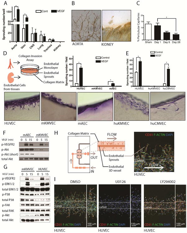
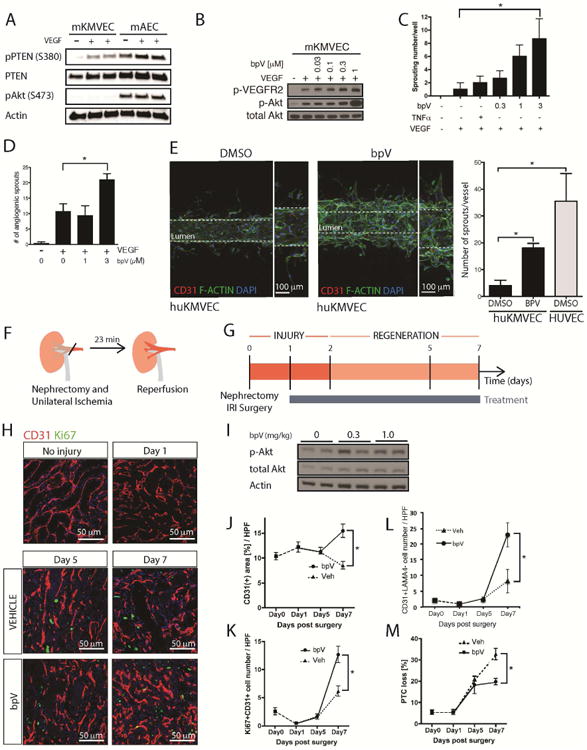
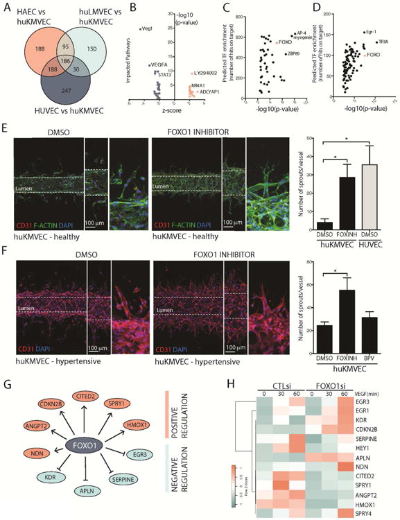
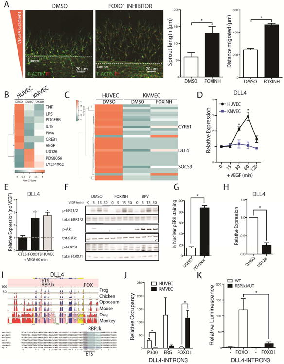
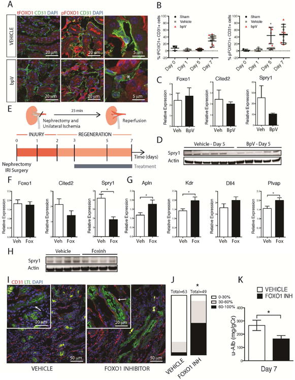
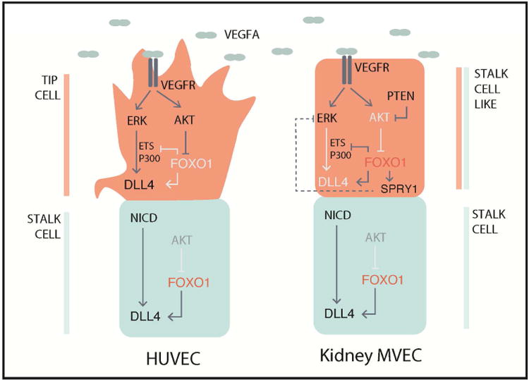
Loss of the microvascular (MV) network results in tissue ischemia, loss of tissue function, and is a hallmark of chronic diseases. The incorporation of a functional vascular network with that of the host remains a challenge to utilizing engineered tissues in clinically relevant therapies. We showed that vascular-bed-specific endothelial cells (ECs) exhibit differing angiogenic capacities, with kidney microvascular endothelial cells (MVECs) being the most deficient, and sought to explore the underlying mechanism. Constitutive activation of the phosphatase PTEN in kidney MVECs resulted in impaired PI3K/AKT activity in response to vascular endothelial growth factor (VEGF). Suppression of PTEN in vivo resulted in microvascular regeneration, but was insufficient to improve tissue function. Promoter analysis of the differentially regulated genes in KMVECs suggests that the transcription factor FOXO1 is highly active and RNAseq analysis revealed that hyperactive FOXO1 inhibits VEGF-Notch-dependent tip-cell formation by direct and indirect inhibition of DLL4 expression in response to VEGF. Inhibition of FOXO1 enhanced angiogenesis in human bio-engineered capillaries, and resulted in microvascular regeneration and improved function in mouse models of injury-repair.

Keywords: Angiogenesis; FOXO1; Microfluidics; VEGF; Vascular rarefaction; Vascular regeneration.


Conflict of interest statement

Conflicts of Interest: Employees of Biogen have company stock. J.S.D has patents for therapies to stimulate organ regeneration.

Figures

Fig 1
Fig 1. Deficient angiogenesis from solid organ endothelium is associated with attenuated ERK and AKT signaling
(A) Graph of angiogenic sprouts 72h after placing 1 mm3 pieces of internal organs in collagen gel in the presence or absence of 50ng/ml VEGF-A. (B) Light micrographs showing CD31-stained (brown) new vessels sprouting from aorta but not from kidney (C) Graph showing the % area of peritubular capillaries in the kidney following IRI. (D-E) Schema, toluidine blue stained thick sections and graphs showing angiogenesis in a gel invasion assay of mouse (D) or human (E) endothelial cells. Note MVECs from both mouse and human kidney showed very little capacity to form angiogenic sprouts and this was not enhanced by the addition of VEGF-A. (F) Blots showing activated (phosphorylated) VEGFR2, AKT in mAECs compared to m KMVECs. Note that KMVECs show very little activated AKT. (G) Bots showing activated VEGFR2, ERK, P38, FAK and AKT comparing mouse KMVECs and HUVECs. Note KMVECs have attenuated p-ERK1/2, and almost absent p-AKT in response to VEGF. (H) Schema and image of angiogenesis in human capillaries microfabricated in microfluidic devices. (I) Images of human capillaries generated from HUVECs under flow, 4 days after stimulating the vessels with VEGF-A in the surrounding gel and lumen to induce angiogenesis. Note widespread formation of stalks into the gel and the presence of tip cells with filopodia in the presence of vehicle (DMSO). However, the in the presence of the MEK inhibitor U0126, HUVEC cells migrate away from the vessel into the surrounding matrix, however, they are unable to form connections to the parent vessel or tip-and-stalk structures. In the presence of LY294002, which inhibits PI3K, new capillary formation is markedly attenuated with individual cells migrating out of the vessel wall similar to but less extensive than the effect of U0126. (n = 5 per group; P < 0.05)
Fig 2
Fig 2. High levels of active PTEN in MVECs can be overcome by the inhibitor bpV to restore angiogenesis and stimulate microvascular regeneration
(A) Blots showing levels of total and inactive (phosphorylated) PTEN as well as active AKT. KMVECs have higher levels of active PTEN. (B) Blots showing the effect of concentrations of the PTEN inhibitor bpV on activate AKT in response to VEGF in mouse ECs. (C-D) bpV permits successful angiogenesis in the gel invasion assay of mouse (C) and human (D) MVECs from kidney. (E) In microfluidic devices under flow human kidney MVECs do not undergo sprouting angiogenesis in response to VEGF-A (left panel), rather individual cells migrate from the capillary, but are not connected to the lumen wall. By contrast (right) in the presence of bpV, successful angiogenesis occurs with sprouts that are connected to the parent vessel. In direct comparison bpV permits KMVECs to form new capillary sprouts at approximately half the level shown by HUVECs. (F-G) Schema and timecourse showing injury/repair caused by IRI in mouse kidney, the phase of regeneration and the duration of treatment with bpV. (H) Representative fluorescence images of kidneys after IRI, from mice treated with vehicle or bpV (I) Western blots of whole kidneys showing bpV effect on P-AKT (J-K) Effect of bpV on CD31 area (J) and proliferating ECs (K) in whole tissue sections (L) Effect of bpV on tip cell formation as defined by EC sprout without associated basal lamina detected by lamanina4 (M) Effect of bpV on total capillary loss. (n = 5-7 animals per group; P < 0.05).
Fig 3
Fig 3. KMVECs are characterized by enhanced FOXO1 activity that can be inhibited to restore angiogenesis in functioning human kidney capillaries
(A) Venn diagram showing overlapping and unique genes expressed by different types of endothelial cells. (B) Upstream analysis of the 186 genes enriched in KMVECs compared to HAECs showing genes consistent with impaired VEGF signaling as well as STAT3 signaling, and expression of genes consistent with blockade of PI3K/AKT by LY924002. (C-D) Transcription factor binding to promoters of DEGs unique to KMVECs (C) and unique to VEGF regulated genes identified in B (D) shows a large number of conserved FOXO1 binding sites at these promoters, thus pointing to enhanced FOXO1 activity (E-F) Images and results from microfluidic devices containing human kidney capillaries from young healthy kidneys (n=6 per group, 2 donors) (E) or aged hypertensive kidneys (n=5 per group, 1 donor) (F), stimulated to undergo sprouting angiogenesis with VEGF. In the presence of the FOXO1 inhibitor AS1842856 both healthy capillaries and aged capillaries successfully form new capillaries with tip-stalk structures. FOXO1 inhibition enables angiogenic capacity in kidney MVECs like that seen in HUVECs. Capillaries engineered from aged hypertensive kidneys had a higher tendency to form new vascular structures, although these did not show tip cell morphology (F, lower left images). FOXO1 inhibition enhanced new vessel formation with obvious tip cell morphology. Note bpV was less effective in aged capillary. (G) Pathway analysis of the DEGs in KMVECs showed genes predicted to be regulated by FOXO1 (H) Effect of siRNA against FOXO1 in KMVECs on potential FOXO1 target genes from G while under dynamic stimulation by VEGF-A by quantitative RT-PCR. Note CITED2 and SPRY1, identified as novel targets of FOXO1, show marked dynamic regulation by VEGF-A.
Fig 4
Fig 4. FOXO1 suppresses VEGF-A mediated nuclear ERK signaling to prevent activation of the tip cell marker DLL4
(A) Images from a dual channel microfluidic system with a constant lateralized VEGF gradient showing that inhibition of FOXO1 induces greater response to VEGF in KMVECs which results in both increased sprout length and increased distance migrated (B) Heatmap showing upstream pathways, detected by RNA sequencing and analyzed by IPA, hierarchically clustered that are enriched in HUVECs, and KMVECs following 1 hr treatment with FOXO1 inhibitor and 30 mins stimulation with VEGF-A (C) Heatmap showing hierarchical clustering of the genes most enriched in HUVECs and KMVECs following treatment with FOXO1 inhibitor and 30 mins after treatment with VEGF-A. DLL4 is most strongly de-repressed when FOXO1 was inhibited. Other genes include CYR61 and SOCS3 (D) Time course of DLL4 transcription in HUVECs and KMVECs in response to VEGF-A. KMVECs are unresponsive. (E) Knock down of FOXO1 in KMVECs restored activation of DLL4 expression in response to VEGF-A (F) Blots showing bpV enhances p-AKT and p-FOXO1 but neither inhibitor enhances total pERK in MVECs. (G) Quantification of pERK localization within KMVECs following stimulation by VEGF-A for 60 minutes in the presence and absence of FOXO1 inhibitor. pERK is predominantly cytoplasmic in KMVECs after VEGF-A, but in the presence of FOXO1 inhibitor it is predominantly nuclear (H) Q-PCR results showing in expression of DLL4 is reduced when MEK is inhibited. (I) Conserved putative transcription factor binding sites within the DLL4 locus, specifically focusing in on Intron 3 where both ETS and RBPJk sites are 100% conserved between many vertebrates. (J) ChIP of EP300 and ERG show low binding to the DLL4 intron 3 in KMVEC compared to HUVEC with the converse observed with FOXO1 occupancy at this locus. (K) Luciferase assay with a reporter construct containing the Intron 3 of DLL4 as well as the RBPJk mutant version. Overexpression of FOXO1 induces the wild type construct but not the RBPJk mutant (n = 3-6/group; * P < 0.05).
Fig 5
Fig 5. FOXO1 inhibition in vivo enhances angiogenesis, revascularization and organ function during injury/repair
(A-B) Fluorescence images and (B) quantification of total and P-FOXO1 in mouse kidneys after IRI in the presence of bpV or vehicle. Note increased P-FOXO1 in endothelium in the presence of bpV day 5 after IRI (C). Q-PCR of kidneys d5 after IRI showing the effect of bpV on transcript levels (D) Whole tissue western blots showing levels of Sprouty1 in the presence of bpV (E) Schema and timecourse showing injury/repair caused by IRI in mouse kidney, the phase of regeneration and the duration of treatment with FOXO1 inhibitor (F) Q-PCR results showing the effect of FOXO1 inhibition on whole tissue levels of target genes Cited2 and Spry1 at day 5 after injury (G) Q-PCR showing the effect of FOXO1 inhibition on levels of angiogenesis and endothelial markers in kidney day 7 after IRI including the VEGF-A dependent FOXO1 regulated genes, such as Apln, Kdr, and Dll4. (H) Western blot showing whole tissue levels of Sprouty1 d5 after injury (I-J) Fluorescence images and quantification of endothelial density in the kidney following IRI. Note the preservation of capillaries around proximal tubules in kidneys treated with FOXO1 inhibitor. Graph shows proportion of kidney with tubules that have differing levels of capillary coverage (K) Urinary Albumin/Creatinine ratio at 7d after IRI from mice treated with FOXO1 inhibitor compared to vehicle. (n = 5 animals/group, *P < 0.05)
Fig 6
Fig 6. Model incorporating the role of FOXO1 in sprouting angiogenesis
Induction of sprouting angiogenesis traditionally studied in HUVECs (left side) occurs through the activation of the VEGFR by ligand binding, which elicits a signaling cascade to activate DLL4 expression through two major signaling pathways, MAPK/ERK and PI3K/AKT. ERK signaling serves to increase ETS and EP300 recruitment to activate expression of DLL4, and AKT serves to inhibit FOXO1 activity allowing for the recruitment of transcriptional activators. Expression of DLL4 in turn activates Notch signaling in neighboring stalk cells, which has reduced VEGF-induced AKT signaling, resulting in high FOXO1 activity. High FOXO1 activity in the stalk cells serves to maintain DLL4 expression and prevents ETS/EP300 recruitment to Intron 3 of DLL4. Kidney MVECs (right side) however are desensitized to VEGF induced signaling due to hyperactive PTEN thereby inhibiting phosphorylation and activation of AKT, resulting in high FOXO1 activity even in the presence of VEGF. FOXO1 inhibits recruitment of transcriptional activators ETS and EP300 to Intron 3 of DLL4. In addition, FOXO1 may regulate the expression of the ERK inhibitor SPRY1 which may further impact ETS and EP300 activities. Altogether, this results in a lack of tip cell fate and reduced angiogenic capacity.

References

    1. Ye L, et al. Transplantation of nanoparticle transfected skeletal myoblasts overexpressing vascular endothelial growth factor-165 for cardiac repair. Circulation. 2007;116(11 Suppl):I113–20. - PubMed
    1. Kolesky DB, et al. 3D bioprinting of vascularized, heterogeneous cell-laden tissue constructs. Adv Mater. 2014;26(19):3124–30. - PubMed
    1. Kolesky DB, et al. Three-dimensional bioprinting of thick vascularized tissues. Proc Natl Acad Sci U S A. 2016;113(12):3179–84. - PMC - PubMed
    1. Mishra R, et al. Effect of prevascularization on in vivo vascularization of poly(propylene fumarate)/fibrin scaffolds. Biomaterials. 2016;77:255–66. - PMC - PubMed
    1. Brudno Y, et al. Enhancing microvascular formation and vessel maturation through temporal control over multiple pro-angiogenic and pro-maturation factors. Biomaterials. 2013;34(36):9201–9. - PMC - PubMed

MeSH terms

Substances