. 2017 Jul 15;6(7):e005318.
doi: 10.1161/JAHA.116.005318.

Identification of apoB-100 Peptide-Specific CD8+ T Cells in Atherosclerosis

Affiliations

Identification of apoB-100 Peptide-Specific CD8+ T Cells in Atherosclerosis

Paul C Dimayuga et al. J Am Heart Assoc. .

Abstract

Background: T cells are found in atherosclerotic plaques, with evidence supporting a potential role for CD8+ T cells in atherogenesis. Prior studies provide evidence of low-density lipoprotein and apoB-100 reactive T cells, yet specific epitopes relevant to the disease remain to be defined. The current study was undertaken to identify and characterize endogenous, antigen-specific CD8+ T cells in atherosclerosis.

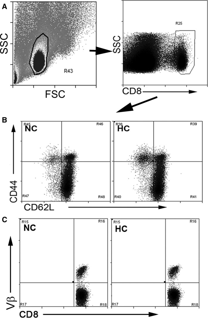
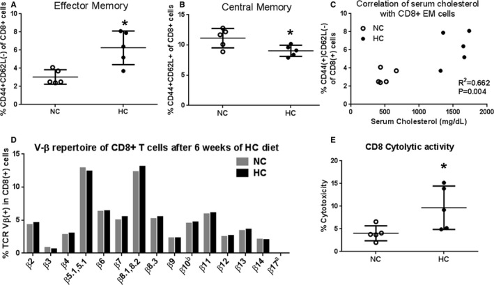
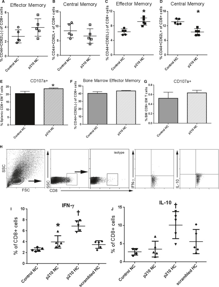
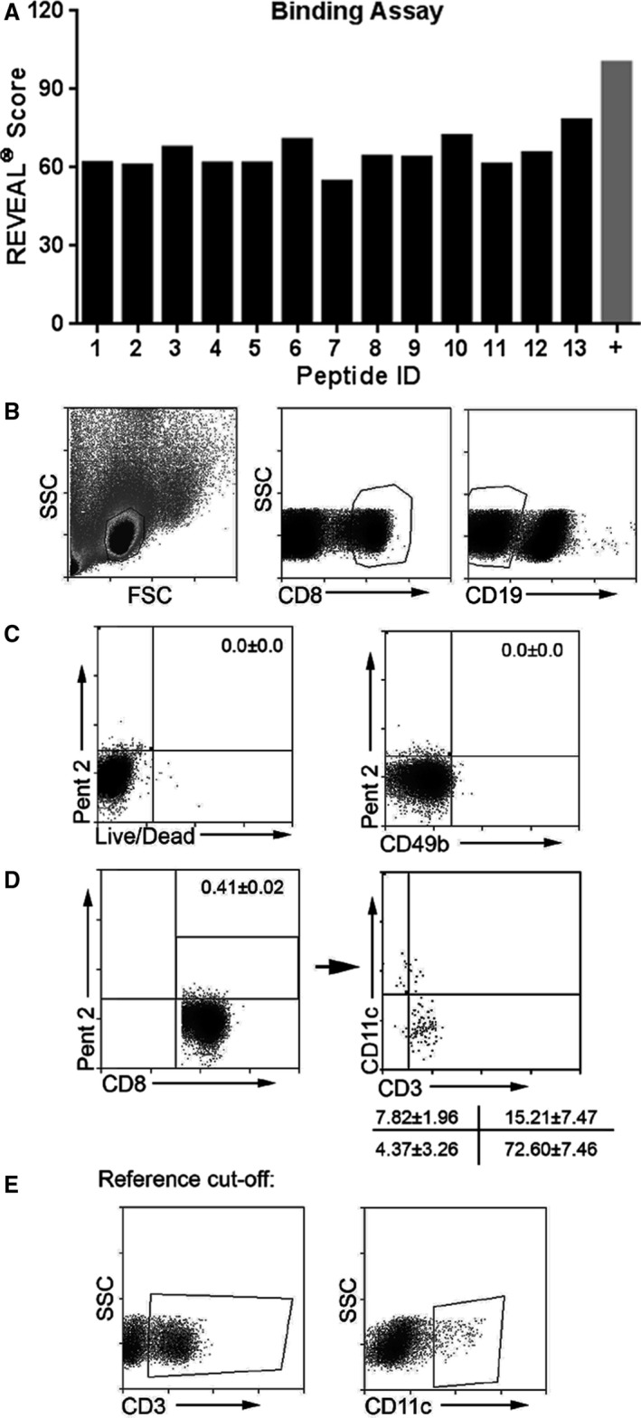
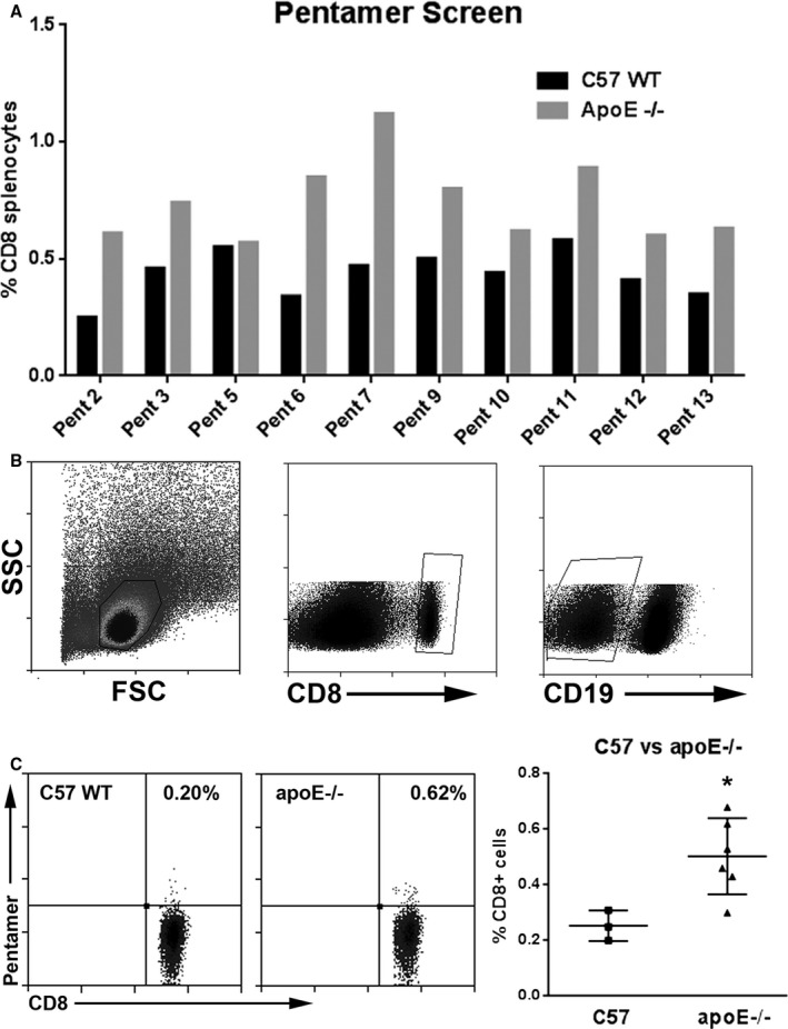
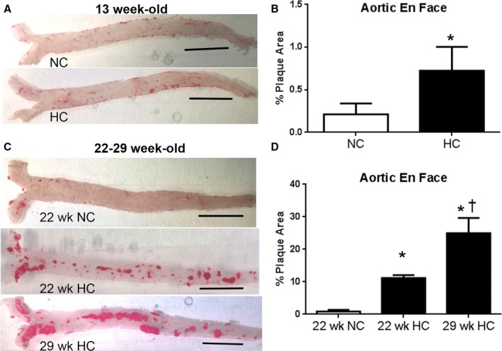
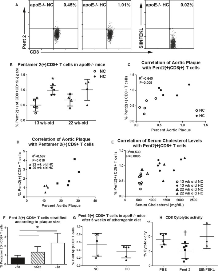
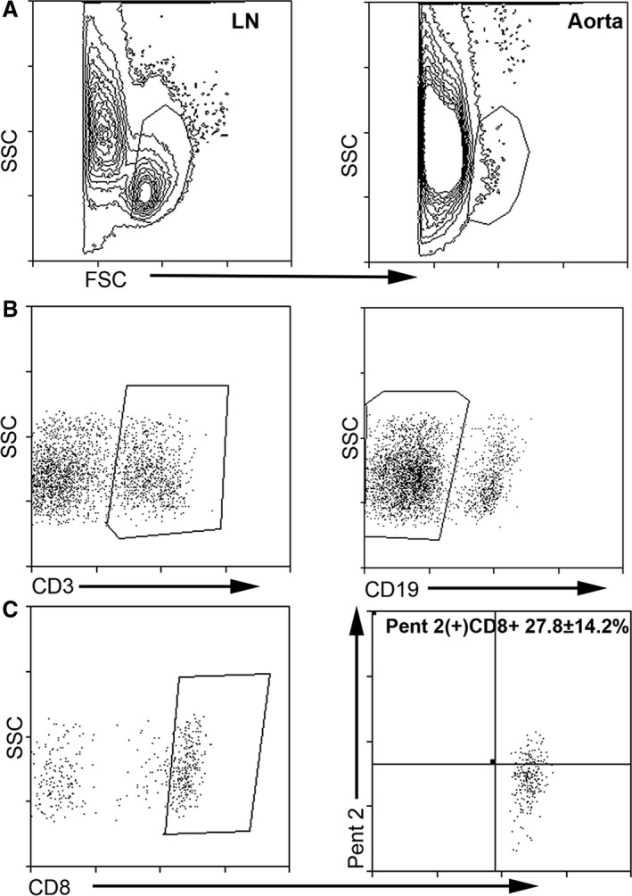
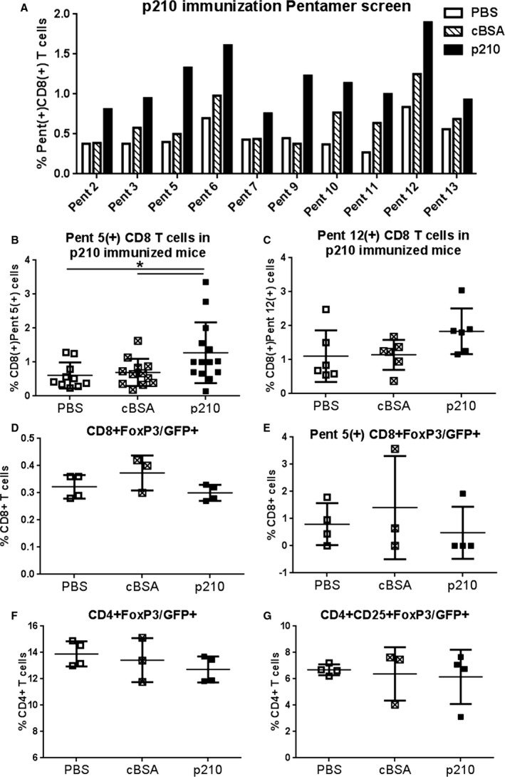
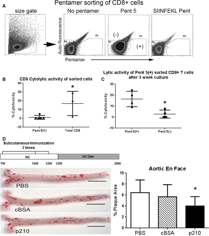
Methods and results: A peptide fragment of apoB-100 that tested positive for binding to the mouse MHC-I allele H2Kb was used to generate a fluorescent-labeled H2Kb pentamer and tested in apoE-/- mice. H2Kb pentamer(+)CD8+ T cells were higher in apoE-/- mice fed an atherogenic diet compared with those fed a normal chow. H2Kb pentamer (+)CD8+ T cells in atherogenic diet-fed mice had significantly increased effector memory phenotype with a shift in Vβ profile. H2Kb pentamer blocked lytic activity of CD8+ T cells from atherogenic diet-fed mice. Immunization of age-matched apoE-/- mice with the apoB-100 peptide altered the immune-dominant epitope of CD8+ T cells and reduced atherosclerosis.

Conclusions: Our study provides evidence of a self-reactive, antigen-specific CD8+ T-cell population in apoE-/- mice. Immune modulation using the peptide antigen reduced atherosclerosis in apoE-/- mice.

Keywords: CD8+ T cells; apoB‐100; atherosclerosis; immunology; lymphocyte; major histocompatibility complex‐I tetramer.


Figures

Figure 1
Figure 1
Gating strategy for CD8+ T cells in apoE−/− mice fed normal chow or an atherogenic diet. Splenocytes from mice fed normal chow (NC) or atherogenic diet (HC) were size gated on lymphocytes and then on CD8 (A). Gated cells were then plotted on CD62L/CD44 (B) for memory cell profile or CD8/Vβ (C) to assess Vβ repertoire. Nonviable cells comprised <0.05% of freshly isolated, stained splenocytes. FSC indicates Forward Scatter; SSC, Side Scatter.
Figure 2
Figure 2
Atherogenic diet–induced generation of effector memory CD8+ T cells and increased cytolytic activity. A, CD8+ effector memory T cells and (B) central memory CD8+ T cells of freshly isolated splenocytes from apoE−/− mice fed normal chow (NC) or an atherogenic diet (HC) for 6 weeks. *P<0.05, N=5 each. Gating strategy is shown in Figure 1. C, CD8+ effector memory T‐cell correlation with serum cholesterol (R 2=0.662; P=0.004). D, Bar graph of Vβ repertoire after 6 weeks of atherogenic diet compared with NC–fed mice. Spleens from 5 mice per group were pooled to obtain a sufficient number of cells for all Vβ types. Gating strategy and representative scatterplot for Vβ staining analysis of splenocytes are shown in Figure 1C. E, Cytolytic activity of CD8+ T cells from apoE−/− mice fed NC or an HC for 6 weeks. *P<0.01; N=5 each.
Figure 3
Figure 3
CD8+ T‐cell response to stimulation with p210 peptide. Splenocytes from apoE−/− mice fed normal chow (NC) or atherogenic diet (HC) for 12 weeks were stimulated for 24 hours and then stained for memory cell profile. A and B, CD8+ effector memory and central memory in splenocytes from NC mice. C and D, CD8+ effector memory and central memory in splenocytes from HC mice. N=5 each; P<0.05. E, CD107a staining, a marker of CD8+ T‐cell degranulation as a function of cytolytic activity, was significantly increased after 5‐hour p210 stimulation in splenic CD8+ effector memory T cells (N=4 each, P<0.05). F, Bone marrow cells had more effector memory cells as a percentage of total CD8+ T cells as compared with splenocytes (C) but did not change after 24‐hour p210 stimulation (N=3 each; P=NS, Mann–Whitney test). CD107a+ staining was also unchanged after 5‐hour p210 stimulation in bone marrow (BM) CD8+ effector memory T cells (G; N=3 each; P=NS, Mann–Whitney test). Intracellular cytokine staining in splenocytes of apoE−/− mice fed NC or HC for 6 weeks (H through J). Splenocytes were stimulated with p210 peptide, scrambled peptide, or vehicle as control for 48 hours, with Brefeldin‐A treatment for the last 4 hours before collection and staining. Cells were size‐gated for lymphocytes and then for CD8 with isotype staining as reference. Representative scatterplots of cells stained for IFN‐γ and IL‐10 (H). CD8+IFN‐γ (I) and CD8+IL‐10+ (J) cells in splenocytes of mice fed NC or HC. N=5 each; *P<0.05 vs Control NC; P<0.05 vs all groups, ANOVA. FSC indicates Forward Scatter; IFN‐γ, interferon‐γ; IL‐10, interleukin‐10; NS, not significant; SSC, Side Scatter.
Figure 4
Figure 4
Screening of p210 peptide fragments for binding on mouse H2Kb. A, REVEAL binding assays identified 13 possible 8‐mer sequences for H2Kb binding and 10 were synthesized successfully as pentamers in the ProVE platform, the first of which was Pent 2 (see Table 2). B through E, Optimization and characterization of pentamer staining and gating strategy were performed using freshly isolated splenocytes from apoE−/− mice with cell viability staining and antibodies to CD3e, CD8, CD11c, CD19, CD49b, and Pent 2; representative of 3. B, Splenocytes were size‐gated and selected for the CD8+CD19(−) gate. C, Cells gated from (B) were plotted on live/dead cell vs Pent 2 stain quadrant analysis (left) or CD49b (NK cells) vs Pent 2 stain quadrant analysis. Numbers in the top right quadrants are percentage of cells double positive for the indicated stains. D, Pent 2(+)CD8+ T cells as plotted (left) after CD8+CD19(−) gating scheme shown in (B). Box indicates double positive cells with corresponding percentage. These cells were then plotted on CD3e vs CD11c for further characterization (right). Numbers at lower right are percentages of the respective cell subgroups corresponding to the plot quadrants. E, CD3e (left) and CD11c (right) positive cells on the SSC plot used as reference for the quadrant analysis in (D). FSC indicates Forward Scatter; SSC, Side Scatter.
Figure 5
Figure 5
Pentamer 2(+)CD8+ cells in apoE−/− mice. A, Bar graph of relevant p210 epitopes screened in freshly isolated splenocytes of mice as detected by pentamer(+)CD8+ T cells. Screening for potential relevant epitopes was performed by staining freshly isolated splenocytes from 1 C57Bl6/J or pooled from 5 apoE−/− mice age‐matched at 13 weeks. Black bars=C57Bl6/J; gray bars=apoE−/−. B, Cells were size‐gated for lymphocytes and for CD8+ CD19(−) stain. C, Representative scatterplot of Pent 2(+)CD8+ T cells in splenocytes of C57 wild‐type (C57 WT, left panel) or apoE−/− (middle panel) mice. Pentamer 2(+)CD8+ T cells in splenocytes (right panel) of C57 WT mice (N=3) compared with apoE−/− mice (N=6). *P<0.05. FSC indicates Forward Scatter; SSC, Side Scatter.
Figure 6
Figure 6
Progression of atherosclerosis after atherogenic diet feeding. A, Representative photos of aortic en face oil‐red‐o staining of plaques in 13‐week‐old mice fed NC or HC. B, Measured percentage plaque area in 13‐week‐old mice fed NC or HC (0.21±0.13% vs 0.72±0.28%, respectively; N=5 each; P<0.01). The study was extended over a longer period of time and included 22‐week‐old mice fed normal chow, 22‐week‐old mice fed an atherogenic diet for 15 weeks, and 29‐week‐old mice fed an atherogenic diet for 22 weeks. C, Representative en face oil red‐o staining of plaques in 22‐ and 29‐week‐old mice fed NC or HC. D, Measured percent plaque area in aorta. *P<0.001 vs 22 weeks NC; P<0.001 vs 22 weeks HC (22‐week‐old normal chow: 0.9±0.5%, N=4; 22‐week‐old atherogenic diet: 11.2±0.9%, N=4; 29‐week‐old atherogenic diet: 24.9±4.7%, N=5; P<0.001, ANOVA). Bar = 0.5 cm in 6A and 6C. HC indicates atherogenic diet; NC, normal chow.
Figure 7
Figure 7
Pentamer 2(+)CD8+ cells in apoE−/− mice. A, Representative scatterplot of Pent 2(+)CD8+ T cells in splenocytes of 13‐week‐old apoE−/− mice fed normal chow (apoE−/− NC) or an atherogenic diet (apoE−/− HC). Specificity of the stain was assured by the use of an H2‐Kb allele‐matched, nonrelevant pentamer with the OVA peptide SIINFEKL. B, Graph of Pent 2(+)CD8+ T cells in splenocytes of apoE−/− mice. N=5 each for 13‐week‐old and N=4 each for 22‐week‐old mice. *P<0.05. Open symbol: normal chow; closed symbol: atherogenic diet. C, Percent aortic plaque area was significantly correlated with percent Pent 2(+)CD8+ T cells in 13‐week‐old mice. D, Plaque area was also correlated with Pent 2(+)CD8+ T cells in older apoE−/− mice. E, There was significant correlation between serum levels of total cholesterol and the percentage of Pent 2(+)CD8+ T cells in the 13‐ and 22‐week‐old mice (R 2=0.539, P=0.0005). F, Pent 2(+) CD8+ T cells plotted according to stratified percent aortic plaque size (x‐axis) from mice fed an atherogenic diet for 13 to 22 weeks. <10 N=14; 10 to 20 N=7; >20 N=7; *P<0.05, ANOVA. G, There was no difference in Pent 5(+)CD8+ T cells in 13‐week‐old mice fed NC or HC. H, Pentamer 2 significantly reduced cytolytic activity of CD8+ T cells from mice fed the atherogenic diet for 6 weeks. PBS and Pent 2 N=6 each; SIINFEKL N=3; P<0.05 vs PBS; Kruskal–Wallis and Dunn's multiple comparison test.
Figure 8
Figure 8
Analysis of TCR Vβ use and Pent2(+)CD8+ T cell subtypes in 13‐week‐old apoE−/− mice fed either normal chow or atherogenic diet for 6 weeks. A, Cells were size‐gated for lymphocytes then CD8+, CD19(−), and Pent 2(+) stain. The OVA H2Kb pentamer SIINFEKL was used as control. Representative scatterplot of Vβ staining of CD8+ T cells as gating reference for Vβ analysis, and representative histogram depicting Vβ positive staining in Pent 2(+)CD8+ gated T cells (B, left panels). Representative scatterplot of CD44 and CD62L staining of CD8+ T‐cell gated splenocytes from 13‐week‐old mice fed an atherogenic diet for 6 weeks as reference for Pent 2(+)CD8+ T‐cell analysis (B, right panel). C, Vβ repertoire of Pent 2(+)CD8+ T cells. D, Pent 2(+)CD8+ gated naïve T cells. E, Pent 2(+)CD8+ gated effector memory T cells. F, Pent 2(+)CD8+ gated central memory T cells. *P<0.05; N=5 per group. FSC indicates Forward Scatter; HC, atherogenic diet; NC, normal chow; SSC, Side Scatter; TCR, T‐cell receptor.
Figure 9
Figure 9
Pent 2(+)CD8+ T cells localize in the atherosclerotic plaques of apoE−/− mice. Aortas from apoE−/− mice fed an atherogenic diet for 22 weeks were subjected to enzymatic digestion and stained with the pentamer. Gating strategy was based on doublet and dead cell exclusion using the viability stain. Size gating was then performed based on lymph node cells after the dump gate exclusion (A). Size‐gated cells were then selected (B) for CD3e+, CD19(−), and CD8+ cells then plotted for Pent 2(+) CD8+ T cells (C). FSC indicates Forward Scatter; LN, lymph node cells; SSC, Side Scatter.
Figure 10
Figure 10
Pentamer(+) CD8+ T cells in apoE−/− mice immunized with the apoB‐100 peptide p210. A, Mice were immunized at 7 weeks of age and administered with booster shots at 10 and 12 weeks of age. At 13 weeks of age, mice were euthanized and the spleens collected for pentamer screening. Splenocytes from 5 mice per group were pooled. B, The observed increase in Pent 5(+)CD8+ T cells in immunized mice was confirmed in individual mice (PBS N=10; cBSA N=12; p210 N=14; *P<0.05). C, Pent 12(+)CD8+ T cells were also tested in individual mice (N=6 each; P=NS). ApoE−/− mice expressing the GFP protein on the FoxP3 promoter immunized with p210 did not result in changes in total CD8+FoxP3+ (D) or in Pent 5(+)CD8+FoxP3+ T cells (E). No differences were observed in CD4+FoxP3+ or CD4+CD25+FoxP3+ Treg cells (F and G, respectively); PBS N=4; cBSA N=3; p210 N=4). cBSA indicates cationic bovine serum albumin; NS, not significant.
Figure 11
Figure 11
Functional profile of Pent 5(+)CD8+ T cells and atherosclerosis in apoE−/− mice immunized with the apoB‐100 peptide p210. A, Negatively isolated CD8+ T cells pooled from 5 mice immunized with p210 were sorted based on Pent 5 staining using unstained cells and SIINFEKL pentamer‐stained cells as reference. B, Sorted CD8+ T cells were then used in a 4‐hour cytolytic assay with dendritic cells as targets. Pent 5(+) N=5; total CD8 N=3; P<0.05. C, An aliquot of the sorted cells was further cultured for 21 days with IL‐2 supplementation (see Methods), collected, and used in the cytolytic assay. N=4 each; P<0.05. D, Immunization with p210 according to the illustrated schedule reduced aortic atherosclerosis in apoE−/− mice at 25 weeks of age as measured by oil red‐o stained lipid area. Representative photos of en face oil red‐o stain of aortas (bar=0.5 cm). Measurements of percentage plaque area were plotted. PBS N=22; cBSA N=21; p210 N=25; *P<0.01 vs PBS and cBSA. cBSA indicates cationic bovine serum albumin; HC, atherogenic diet; IL‐2, interleukin‐2; NC, normal chow.

References

    1. Koltsova EK, Garcia Z, Chodaczek G, Landau M, McArdle S, Scott SR, von Vietinghoff S, Galkina E, Miller YI, Acton ST, Ley K. Dynamic T cell‐APC interactions sustain chronic inflammation in atherosclerosis. J Clin Invest. 2012;122:3114–3126. - PMC - PubMed
    1. Dimayuga PC, Chyu KY, Kirzner J, Yano J, Zhao X, Zhou J, Shah PK, Cercek B. Enhanced neointima formation following arterial injury in immune deficient Rag‐1‐/‐ mice is attenuated by adoptive transfer of CD8 T cells. PLoS One. 2011;6:e20214. - PMC - PubMed
    1. Dimayuga PC, Chyu KY, Lio WM, Zhao X, Yano J, Zhou J, Honjo T, Shah PK, Cercek B. Reduced neointima formation after arterial injury in CD4‐/‐ mice is mediated by CD8+CD28hi T cells. J Am Heart Assoc. 2013;2:e000155 DOI: 10.1161/JAHA.113.000155. - DOI - PMC - PubMed
    1. Chyu KY, Zhao X, Dimayuga PC, Zhou J, Li X, Yano J, Lio WM, Chan LF, Kirzner J, Trinidad P, Cercek B, Shah PK. CD8 T cells mediate the athero‐protective effect of immunization with an ApoB‐100 peptide. PLoS One. 2012;7:e30780. - PMC - PubMed
    1. Kyaw T, Winship A, Tay C, Kanellakis P, Hosseini H, Cao A, Li P, Tipping P, Bobik A, Toh BH. Cytotoxic and proinflammatory CD8+ T lymphocytes promote development of vulnerable atherosclerotic plaques in apoE‐deficient mice. Circulation. 2013;127:1028–1039. - PubMed

MeSH terms