. 2017 Oct 20;27(12):874-911.
doi: 10.1089/ars.2017.7140. Epub 2017 Sep 8.

Regulation of Cellular Redox Signaling by Matricellular Proteins in Vascular Biology, Immunology, and Cancer

Affiliations

Regulation of Cellular Redox Signaling by Matricellular Proteins in Vascular Biology, Immunology, and Cancer

David D Roberts et al. Antioxid Redox Signal. .

Abstract

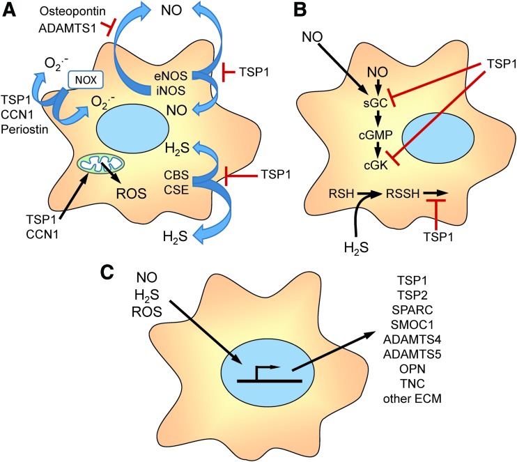
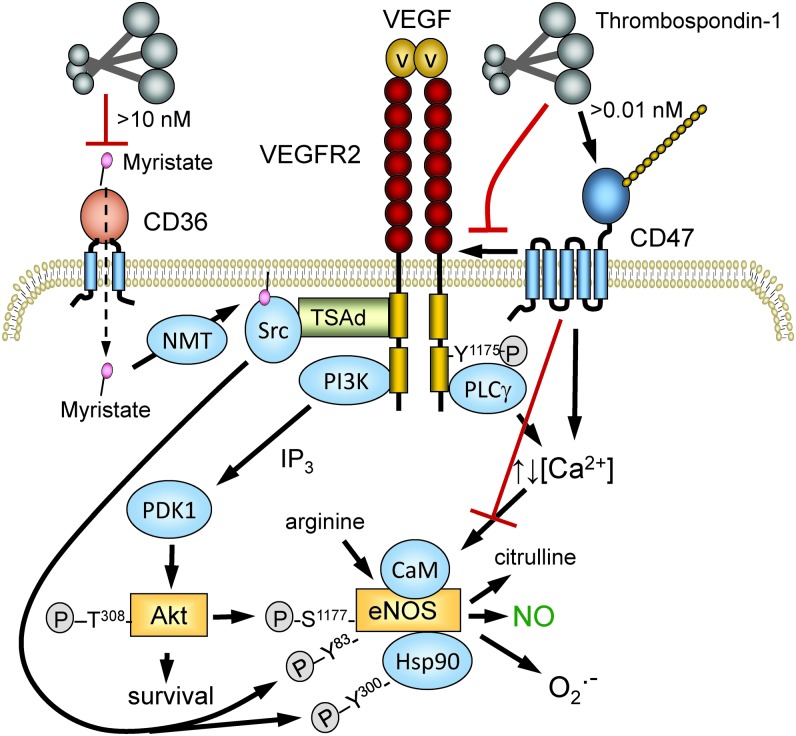
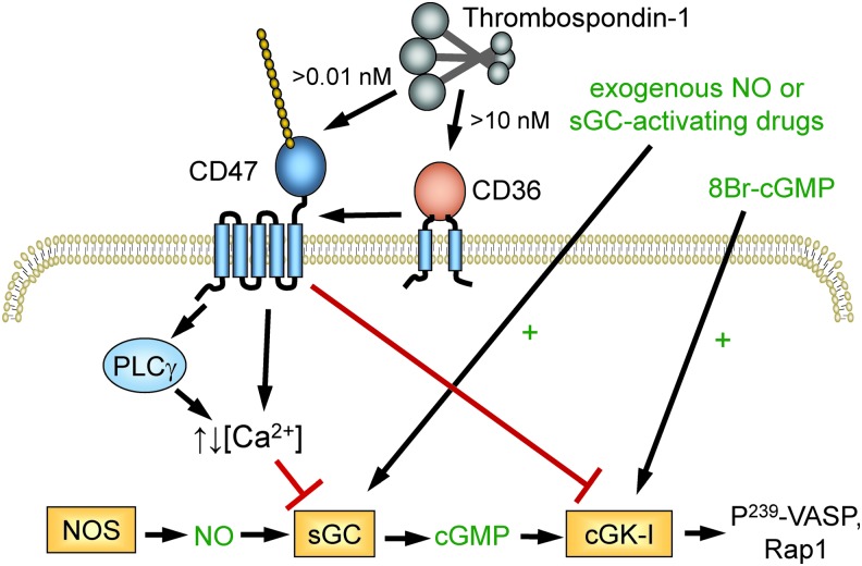
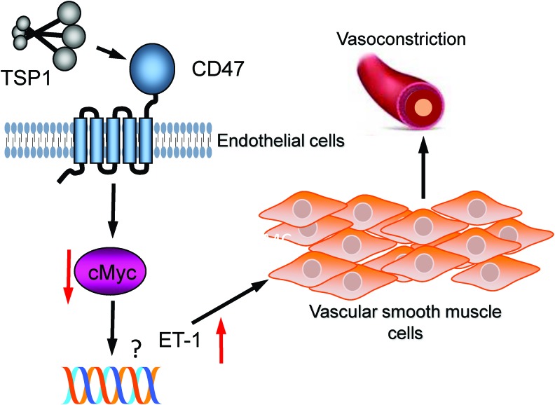
Significance: In contrast to structural elements of the extracellular matrix, matricellular proteins appear transiently during development and injury responses, but their sustained expression can contribute to chronic disease. Through interactions with other matrix components and specific cell surface receptors, matricellular proteins regulate multiple signaling pathways, including those mediated by reactive oxygen and nitrogen species and H2S. Dysregulation of matricellular proteins contributes to the pathogenesis of vascular diseases and cancer. Defining the molecular mechanisms and receptors involved is revealing new therapeutic opportunities. Recent Advances: Thrombospondin-1 (TSP1) regulates NO, H2S, and superoxide production and signaling in several cell types. The TSP1 receptor CD47 plays a central role in inhibition of NO signaling, but other TSP1 receptors also modulate redox signaling. The matricellular protein CCN1 engages some of the same receptors to regulate redox signaling, and ADAMTS1 regulates NO signaling in Marfan syndrome. In addition to mediating matricellular protein signaling, redox signaling is emerging as an important pathway that controls the expression of several matricellular proteins.

Critical issues: Redox signaling remains unexplored for many matricellular proteins. Their interactions with multiple cellular receptors remains an obstacle to defining signaling mechanisms, but improved transgenic models could overcome this barrier.

Future directions: Therapeutics targeting the TSP1 receptor CD47 may have beneficial effects for treating cardiovascular disease and cancer and have recently entered clinical trials. Biomarkers are needed to assess their effects on redox signaling in patients and to evaluate how these contribute to their therapeutic efficacy and potential side effects. Antioxid. Redox Signal. 27, 874-911.

Keywords: CD47; hydrogen sulfide; matricellular proteins; nitric oxide; reactive oxygen species; thrombospondin-1.


Conflict of interest statement

J.S.I. serves as Chair of the Scientific Advisory Board of Radiation Control Technologies, Inc. (RCTI, Garden City, NJ) and has equity interest in RCTI and Tioma Therapeutics (St. Louis, MO) that have licensed CD47 technology for development. The other authors have no competing financial interests to disclose.

Figures

<b>FIG. 1.</b>
FIG. 1.
Matricellular protein families. The thrombospondin family in vertebrates consists of five members. TSP1 and TSP2 are trimeric proteins linked through disulfides in the coiled-coil region that follows the N-terminal HBDs. TSP1 and TSP2 contain three TSRs, which are absent in TSP3, TSP4, and COMP. The latter constitute a second subfamily of homopentamers linked through their coiled-coil regions. A HBD is absent in COMP. The SPARC family consists of SPARC, SPARC-like 1/Hevin, two SPARC-related modular calcium binding proteins (SMOC1 and SMOC2), follistatin-like 1, and three PARC/osteonectin, cwcv, and kazal-like domains proteoglycan proteins (SPOCK1–3). The follistatin and acidic domains are conserved among the family members, but the EF-hand of SPARC is not conserved. The CCN family consists of six members: CCN1, CYR61 (cysteine-rich angiogenic protein 61); CCN2, CTGF; CCN3, NOV; CCN4, WISP1 (WNT1-inducible signaling pathway protein-1); CCN5, WISP2 (WNT1-inducible signaling pathway protein-2); and CCN6, WISP3 (WNT1-inducible signaling pathway protein-3). Tenascins are a family of hexameric proteins consisting of tenascin-C, tenascin-R, tenascin-X, and tenascin-W. The number of EGF-like repeats and fibronectin type 3 repeats are variable. The ADAMTS family contains 19 members with variable number of TSRs. ADAMTS, a disintegrin and metalloproteinase with thrombospondin motifs; CCN, CYR61, CTGF (connective tissue growth factor), and NOV (nephroblastoma overexpressed gene); COMP, cartilage oligomeric matrix protein; EF-hand, helix-loop-helix calcium-binding domain; EGF, epidermal growth factor; HBDs, heparin-binding domains; SPARC, secreted protein acidic and rich in cysteine; TSR, type 1 thrombospondin repeat. To see this illustration in color, the reader is referred to the web version of this article at www.liebertpub.com/ars
<b>FIG. 2.</b>
FIG. 2.
Overview of matricellular protein functions in cellular redox signaling. (A) Several matricellular proteins regulate the biosynthesis of redox molecules that mediate cell autonomous and paracellular signaling. TSP1 limits the activation of eNOS. TSP1, CCN1, and periostin regulate the activity of NADPH oxidases (Nox) that produce intracellular and extracellular superoxide (O2•−). TSP1 and CCN1 also regulate mitochondrial production of ROS. TSP1 regulates the biosynthesis of H2S by controlling the expression of CBS and CSE. ADAMTS1 and osteopontin regulate NO production by controlling the expression of iNOS. (B) TSP1 regulates cellular responses to endogenous or exogenous NO by inhibiting the activation of sGC and cGK and cellular responses to H2S by inhibiting known targets of protein sulfhydration and others that remain to be identified. (C) Redox signaling, in turn, regulates the gene expression, post-translational modification, secretion, and extracellular interactions of the indicated matricellular proteins and other extracellular matrix proteins. CBS, cystathionine β-synthase; cGMP; cGK, cGMP-dependent protein kinase; CSE, cystathionine γ-lyase; eNOS, endothelial nitric oxide synthase; iNOS, inducible nitric oxide synthase; ROS, reactive oxygen species; sGC, soluble guanylate cyclase. To see this illustration in color, the reader is referred to the web version of this article at www.liebertpub.com/ars
<b>FIG. 3.</b>
FIG. 3.
Thrombospondin-1 structural domains and receptor binding sites. TSP1 is a homotrimer of ∼150 kDa subunits linked by interchain disulfide bonds. In each folded TSP1 subunit, the Ca-binding repeats wrap around the C-terminal G domain to form the signature domain of TSP1. Binding domains for TSP1 receptors that regulate redox signaling are indicated. To see this illustration in color, the reader is referred to the web version of this article at www.liebertpub.com/ars
<b>FIG. 4.</b>
FIG. 4.
TSP1 regulation of NO synthesis. TSP1 binding to its receptor CD47 on the plasma membrane transduces signals by dissociating its lateral interaction with VEGFR2, by altering cytoplasmic calcium, and by other undefined pathways. Signaling downstream of VEGFR2 through Src and the PI3-kinase/Akt pathway controls the phosphorylation of eNOS at several sites and the phosphorylation of HSP90 associated with eNOS. TSP1, via CD47, also limits eNOS activation separate from effects mediated through VEGFR2. Altered cytoplasmic calcium regulates the binding of calmodulin, which controls the activity of eNOS and its production of NO versus O2•−. At higher concentrations (>10 nM), TSP1 inhibits CD36-mediated uptake of myristic acid (87), limiting substrate for NMT to acylate Src. This modification is necessary for optimal tyrosine phosphorylation of eNOS. NMT, N-myristoyl transferase; VEGFR2, vascular endothelial growth factor receptor-2. To see this illustration in color, the reader is referred to the web version of this article at www.liebertpub.com/ars
<b>FIG. 5.</b>
FIG. 5.
TSP1 regulation of cellular responses to NO. TSP1 binding to CD47 redundantly inhibits signaling induced by endogenous or exogenous NO at the level of sGC and inhibits cGMP-mediated activation of cGK. In platelets, this limits phosphorylation of VASP and Rap1-mediated integrin activation. Picomolar concentrations of TSP1 are sufficient to inhibit sGC via CD47 in vascular cells, whereas >10 nM TSP1 can engage CD36 to inhibit sGC activation in a CD47-dependent manner (92). VASP, vasodilator-stimulated phosphoprotein. To see this illustration in color, the reader is referred to the web version of this article at www.liebertpub.com/ars
<b>FIG. 6.</b>
FIG. 6.
ADAMTS1 regulation of iNOS. Mutation of fibulin-1 (Fbn1) that causes Marfan syndrome results in decreased expression of ADAMTS1 (186). ADAMTS1 cleaves proteins including SDC4 that regulate the induction of NOS2 via an Akt-NF-κB. The resulting production of NO increases proteolytic cleavage of elastin via MMP9, and the loss of elastic fibers in the aorta leads to formation of aneurysms. MMP9, matrix metalloproteinse-9; SDC4, syndecan-4. To see this illustration in color, the reader is referred to the web version of this article at www.liebertpub.com/ars
<b>FIG. 7.</b>
FIG. 7.
TSP1/CD47 signaling in T cells. Presentation of antigen in the context of MHC induces the formation of the immunological synapse and activation of TCR signaling through mediators, including LAT and ZAP70. CD47 signaling acts downstream of these mediators to limit the induction of multiple genes involved in T cell activation and effector function. In addition to cell autonomous inhibition, CD47 signaling inhibits expression of genes encoding the T cell cytokine IL-2 and the α-subunit of the IL-2 receptor (CD25). CD47 also limits activation-dependent induction of the H2S biosynthetic enzymes CBS and CSE. In T cells their activity is required for reorientation of the MTOC, which is required for immune synapse formation. In cytotoxic CD8+ T cells, CD47 regulates expression of the effector GZMB, which mediates antigen-dependent killing of tumor cells. GZMB, granzyme B; IL, interleukin; MHC, major histocompatibility complex; MTOC, microtubule-organizing center; TCR, T cell receptor. To see this illustration in color, the reader is referred to the web version of this article at www.liebertpub.com/ars
<b>FIG. 8.</b>
FIG. 8.
Regulation of osteopontin expression by H2S. (A) Studies in osteoblasts identified CSE as the major source of endogenous H2S biosynthesis mediating induction of OPN, BMP2, and OCN. BMP2 induces these genes via SMAD signaling that activates the transcription factor RUNX2, but this induction is impaired if H2S synthesis is inhibited. Cys123 and Cys132 were identified as sulfhydration targets on RUNX2 that mediate this activity. (B) In bone marrow mesenchymal stem cells, H2S sulfhydrates Cys residues on the calcium channels TRPV3, TRPV6, and TRPM4. This modification controls calcium influx, which controls transcriptional activation of the RUNX2 gene via PKC and the MAP kinase ERK1/2. BMP2, bone morphogenetic protein-2; OCN, osteocalcin; OPN, osteopontin; PKC, protein kinase C. To see this illustration in color, the reader is referred to the web version of this article at www.liebertpub.com/ars
<b>FIG. 9.</b>
FIG. 9.
Model for TSP1 modulation of SIRPα-mediated ROS and NO production. Binding of CD47 to the extracellular V-domain of SIRPα induces tyrosine phosphorylation of the cytoplasmic domain, which recruits the phosphatase SHP2. Binding of its counter-receptor also increases JAK2/STAT signaling to induce NOS2 expression and PI-3-kinase-dependent Rac1 recruitment to Nox1, which activates it to produce extracellular superoxide. Because TSP1 inhibits the interaction of the extracellular domain of SIRPα to CD47 in biochemical assays (84), TSP1 has the potential to inhibit these SIRPα signaling pathways independent of its CD47-mediated modulation of redox signaling. SIRPα, signal regulatory protein α. To see this illustration in color, the reader is referred to the web version of this article at www.liebertpub.com/ars
<b>FIG. 10.</b>
FIG. 10.
TSP1 via CD47 and SIRPα stimulates increased superoxide production. TSP1 (2.2 nM), a concentration found in human plasma in disease, interacts with cell receptors CD47 as well as SIRP-α to stimulate increased superoxide levels. The Nox assembly subunit p47phox is activated (phosphorylated) in cells treated with TSP1 concurrent with increased superoxide. Suppressing Nox1, or interfering with TSP1 interactions with CD47 and SIRP-α, abrogates TSP1-mediated increases in superoxide. In hypoxic endothelial cells TSP1 via CD47 dysregulates caveolin 1 (Cav-1) and eNOS, and is associated with increased superoxide. Superoxide may in a feedback manner interact with NO and sGC to limit NO signaling, and in a feed forward manner to possibly stimulate TSP1 expression. Together these pathways function to limit arterial vasodilation and impede blood flow. To see this illustration in color, the reader is referred to the web version of this article at www.liebertpub.com/ars
<b>FIG. 11.</b>
FIG. 11.
Pulmonary TSP1-CD47 signaling regulates vasoconstriction via endothelin-1. Pulmonary endothelial cells express ET-1 that alters pulmonary tone acutely and stimulates vasculature remodeling chronically. Smooth muscle cells are paracrine targets of ET-1, whereas in pulmonary hypertension ET-1 receptors are targets for current human therapies. Under hypoxic stress, TSP1 acts via CD47 to repress pulmonary endothelial cMyc via mechanism yet to be defined, thus derepressing ET-1 and promoting arterial vasoconstriction. Conversely, blockade of TSP1-CD47 signaling elevates pulmonary cMyc and decreases ET-1 (not shown). ET-1, endothelin-1. To see this illustration in color, the reader is referred to the web version of this article at www.liebertpub.com/ars
<b>FIG. 12.</b>
FIG. 12.
Regulation of ROS signaling by CCN1. CCN1 signaling mediated by binding of its C-terminal domain to α6β1 integrin and a cell surface HSPG increases ROS production via Rac-1-mediated activation of Nox, nSMase-dependent activation of 5Lox, and activation of mitochondrial ROS production. 5Lox, 5-lipoxygenase; HSPG, heparan sulfate proteoglycan; nSMase, neutral sphingomyelinase. To see this illustration in color, the reader is referred to the web version of this article at www.liebertpub.com/ars
<b>FIG. 13.</b>
FIG. 13.
Radioprotection by loss/blockade of CD47 and ROS. Genotoxic stress caused by ionizing radiation results in opposing global metabolic responses in cells that express or lack expression of CD47 (165). Irradiated WT cells lose capacity for glucose uptake associated with loss of the glucose transporter Glut1. Decreased flux through glycolysis, the TCA cycle, and the PPP lead to impaired anabolic pathways required to repair free radical damage of critical cell macromolecules and loss of glutathione and glyoxalase pathway scavenging of reactive carbonyl metabolites generated by radiation-induced free radicals. All of these repair pathways are preserved in CD47-deficient cells. In addition, PGC1α-dependent support of mitochondrial function and numbers is enhanced in the absence of CD47 and minimizes mitochondrial production of ROS (56). PPP, pentose phosphate pathway; TCA, tricarboxylic acid cycle; WT, wild type. To see this illustration in color, the reader is referred to the web version of this article at www.liebertpub.com/ars
<b>FIG. 14.</b>
FIG. 14.
Regulation of stem cell self-renewal and differentiation by TSP1/CD47 and redox signaling. (A) Loss of TSP1 or CD47 induces spontaneous conversion of differentiated cells to a stem cell state with enhanced expression of the stem cell transcription factors cMyc, Sox2, Oct4, and Klf4 and an increased frequency of asymmetric cell division (113). Conversely, increased CD47 expression or exposure to the ligand TSP1 suppresses stem cells. (B) Consistent with the increased NO signaling in Cd47−/− and Thbs1−/− cells, NO donors promote stem cell self-renewal, and NOS inhibitors promote differentiation (12). (C) In vivo, Cd47−/− mice are protected from renal ischemia/reperfusion injury and exhibit increased expression of the stem cell transcription factors (217). Conversely, loss of stem cell transcription factors and self-renewal in injured WT mice can be prevented by treating with a CD47 antibody that blocks TSP1 binding. However, pan-suppression of NOS activity oral L-NAME does not decrease the enhanced transcription factor expression in kidneys in Cd47−/− mice at baseline or after ischemia-reperfusion injury. Therefore, the preservation of self-renewal in Cd47−/− mice is probably independent of enhanced NO biosynthesis. L-NAME, l-NG-nitroarginine methyl ester. To see this illustration in color, the reader is referred to the web version of this article at www.liebertpub.com/ars
<b>FIG. 15.</b>
FIG. 15.
Network analysis of CD47-dependent gene expression in primary lung endothelial cells. A set of redox pathway-related genes (65) was used to analyze published microarray data comparing WT and Cd47−/− mouse lung endothelial cells (113). Extracted data for genes on this list were subjected to Ingenuity pathway analysis. The canonical pathway for production of nitric oxide and reactive oxygen species gave a z-score −1.342 for CD47 dependence (p-value: 1.74 × 10−5). Upregulated genes in CD47-deficient cells are labeled red and downregulated genes are labeled green. In addition to elements of the NO and ROS pathways, this analysis identifies several protein phosphatases as potential transcriptional targets of CD47 signaling, which could also be regulated post-translationally by CD47-dependent ROS and sulfhydration. To see this illustration in color, the reader is referred to the web version of this article at www.liebertpub.com/ars

References

    1. Abais JM, Xia M, Zhang Y, Boini KM, and Li PL. Redox regulation of NLRP3 inflammasomes: ROS as trigger or effector? Antioxid Redox Signal 22: 1111–1129, 2015 - PMC - PubMed
    1. Adams JC. and Lawler J. The thrombospondins. Cold Spring Harb Perspect Biol 3: a009712, 2011 - PMC - PubMed
    1. Agah A, Kyriakides TR, Lawler J, and Bornstein P. The lack of thrombospondin-1 (TSP1) dictates the course of wound healing in double-TSP1/TSP2-null mice. Am J Pathol 161: 831–839, 2002 - PMC - PubMed
    1. Aird AL, Nevitt CD, Christian K, Williams SK, Hoying JB, and LeBlanc AJ. Adipose-derived stromal vascular fraction cells isolated from old animals exhibit reduced capacity to support the formation of microvascular networks. Exp Gerontol 63: 18–26, 2015 - PMC - PubMed
    1. Alblas J, Honing H, de Lavalette CR, Brown MH, Dijkstra CD, and van den Berg TK. Signal regulatory protein alpha ligation induces macrophage nitric oxide production through JAK/STAT- and phosphatidylinositol 3-kinase/Rac1/NAPDH oxidase/H2O2-dependent pathways. Mol Cell Biol 25: 7181–7192, 2005 - PMC - PubMed

MeSH terms