Case Reports
doi: 10.4103/ams.ams_68_16.
Epidermoid Cyst: Clinical and Surgical Case Report
Affiliations
- PMID: 28713757
- PMCID: PMC5502506
- DOI: 10.4103/ams.ams_68_16
Item in Clipboard
Case Reports
Epidermoid Cyst: Clinical and Surgical Case Report
Ann Maxillofac Surg.
2017 Jan-Jun.
Abstract
The epidermoid cyst is a development cyst, skin, filled with keratin and imprisoned by stratified squamous epithelium similar to skin. They are more frequent in males. Clinically presenting as flabby, prevalent over the face, neck and back. They are usually asymptomatic and its etiology is directly linked to trauma, as well as the imprisonment of epithelial rests during embryonic fusion. This study aims to present a case report of a patient 54 years old, with an epidermal cyst in the face, which was treated surgically.
Keywords: Diagnostics; epidermal cyst; surgery.
Conflict of interest statement
There are no conflicts of interest.
Figures
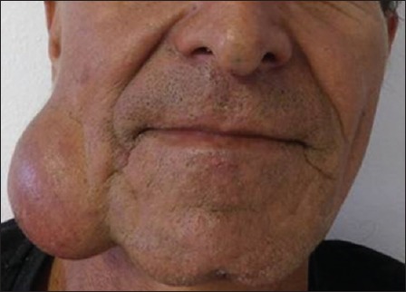
Extensive edema in the right hemiface causing asymmetry from the region of the outer corner of the mouth to the submandibular region
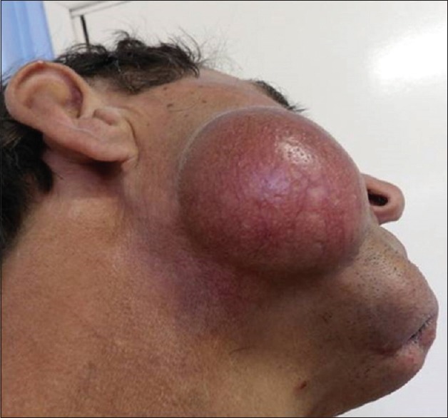
In a lower view, one can better visualize the limits of the lesion. Oval and well delimited
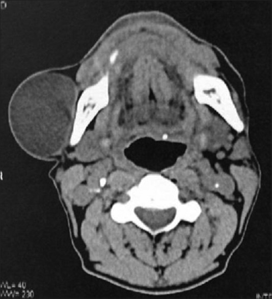
Axial computed tomography. Homogeneous mass, well circumscribed with more than five centimeters in its greatest extensionagreement
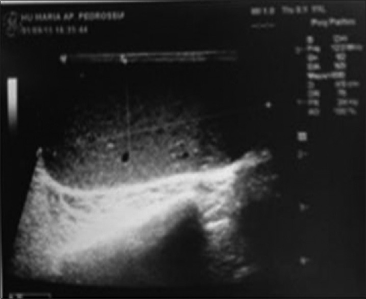
Ultrasound of the cystic lesion. Hypoechoic cystic lesion measuring 5.3 cm × 4.6 cm × 5.1 cm
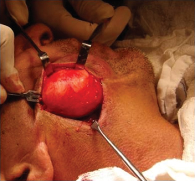
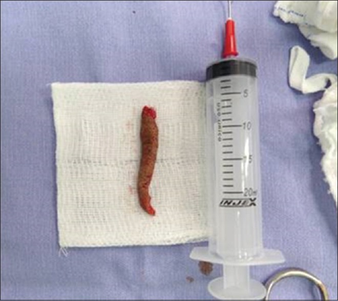
Well delimited mass being removed in a single block, surgically
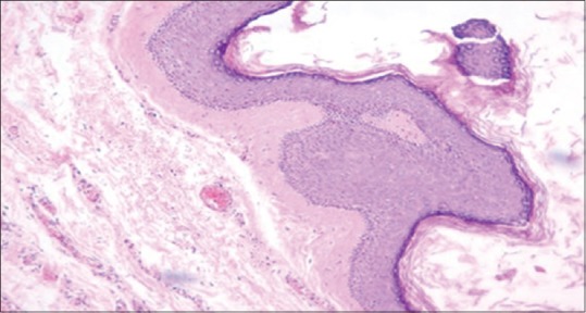
Epidermal cyst-cystic cavity lined by stratified squamous epithelium and filled with corneal lamellae anucleate
Excess skin removed to facilitate primary closure and improvement in esthetic quality after removal of cystic masswithout
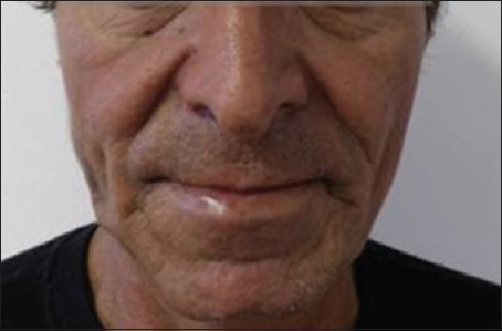
Postoperative appearance of 1 month. Esthetic quality. Facial symmetry
References
-
- Tancredi ARC, Ribeiro-Júnior O, Higo CD, Pedron IG, Lemos-Júnior CA. Epidermoid cyst in the labial comissure region: Case report. Clin Pesq Odontol. 2006;2:329–32.
-
- Kalgutkar A, Kini S, Jambhekar N, Das S. Intradiploic primary epithelial inclusion cyst of the skull. Ann Diagn Pathol. 2006;10:20–3. - PubMed