. 2017 Sep;16(3):3079-3086.
doi: 10.3892/mmr.2017.6997. Epub 2017 Jul 15.

Adipose differentiation‑related protein knockdown inhibits vascular smooth muscle cell proliferation and migration and attenuates neointima formation

Affiliations

Adipose differentiation‑related protein knockdown inhibits vascular smooth muscle cell proliferation and migration and attenuates neointima formation

Haomin Zhao et al. Mol Med Rep. 2017 Sep.

Abstract

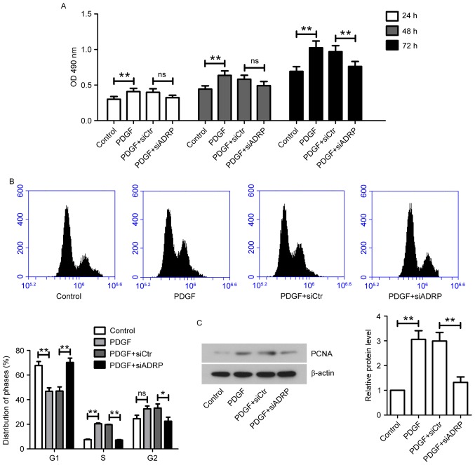
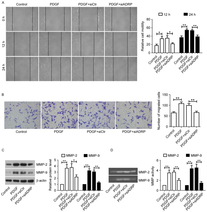
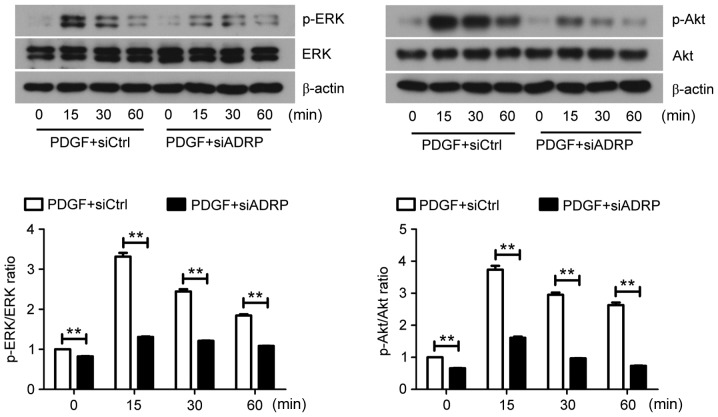
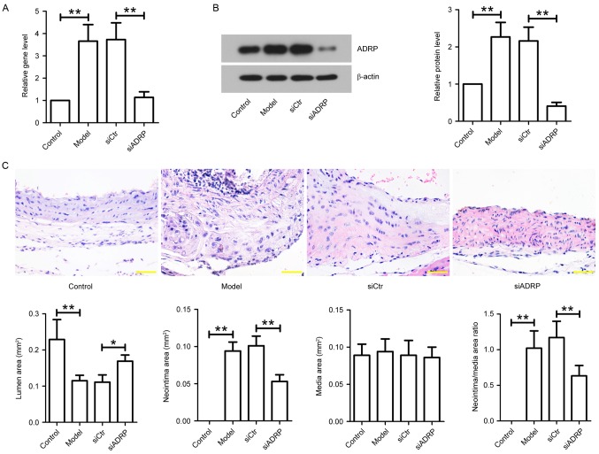
Vascular smooth muscle cells (VSMCs) have an important role in atherosclerosis development. Evidence has demonstrated that adipose differentiation‑related protein (ADRP) is associated with foam cell formation and atherosclerosis progression. However, to the best of our knowledge, no previous studies have investigated the role of ADRP knockdown in platelet‑derived growth factor (PDGF)‑stimulated proliferation and migration of VSMCs in vitro. Furthermore, the effect of ADRP knockdown on neointima formation in vivo remains unclear. In the present study, primary human aortic VSMCs were incubated with PDGF following ADRP small interfering (si)RNA transfection. Cell viability, migration and cell cycle distribution were analyzed by MTT, wound healing and Transwell assays and flow cytometry, respectively. Extracellular signal‑regulated kinase (ERK), phosphorylated (p)‑ERK, Akt, p‑Akt, proliferating cell nuclear antigen (PCNA), matrix metalloproteinase (MMP)‑2 and MMP‑9 protein levels were determined by western blotting. Apolipoprotein E-/- mice fed an atherogenic diet were injected with siADRP or control siRNA twice a week. After 3 weeks of therapy, aortas were excised and ADRP mRNA and protein expression was determined. Neointima formation was assessed by hematoxylin and eosin staining. The results of the current study demonstrated that ADRP knockdown significantly inhibited PDGF‑induced increases in VSMC viability, caused G1 phase cell cycle arrest and decreased PCNA expression. Knockdown of ADRP inhibited PDGF‑induced migration of VSMCs by reducing MMP protein expression and activity. In addition, the present study also demonstrated that ADRP knockdown inhibited ERK and Akt signaling pathways in response to PDGF. Furthermore, siADRP administration suppressed neointima formation in the mouse model. The results of the present study indicate that ADRP may be a potential target for the treatment of atherosclerosis.


Figures

Figure 1.
Figure 1.
Effect of ADRP knockdown on VSMC viability and cell cycle progression induced by PDGF. (A) At 24 h after transfection with siCtr or siADRP, VSMCs were stimulated with 10 ng/ml PDGF. Cell viability was measured by MTT assay at 24, 48 and 72 h. The absorbance at 490 nm was measured. VSMCs were incubated for 48 h with PDGF at 24 h post-transfection. Cell cycle distribution was analyzed by (B) flow cytometry and PCNA protein expression was quantified by (C) western blotting. β-actin served as an internal control. Data are presented as the mean ± standard deviation. *P<0.05 and **P<0.01. ADRP, adipose differentiation-related protein; VSMCs, vascular smooth muscle cells; PDGF, platelet-derived growth factor; siRNA, small interfering RNA; siCtr, control siRNA; siADRP, siRNA targeting ADRP; PCNA, proliferating cell nuclear antigen; OD, optical density.
Figure 2.
Figure 2.
Effect of ADRP knockdown on VSMC migration induced by PDGF. (A) Cell migration was determined by wound healing assay at 12 and 24 h (magnification, ×100). (B) Cell migration ability was confirmed by Transwell migration assay (magnification, ×200). VSMCs were incubated for 24 h with PDGF at 24 h post-transfection. The protein levels and activities of MMP-2 and MMP-9 were examined by (C) western blotting and (D) gelatin zymography. Data are presented as the mean ± standard deviation. *P<0.05 and **P<0.01, as indicated by brackets. ADRP, adipose differentiation-related protein; VSMCs, vascular smooth muscle cells; PDGF, platelet-derived growth factor; MMP, matrix metalloproteinase; siRNA, small interfering RNA: siCtr, control siRNA; siADRP, siRNA targeting ADRP.
Figure 3.
Figure 3.
Effect of ADRP knockdown on the ERK and Akt signaling pathways. At 48 h after siRNA transfection, VSMCs were exposed to PDGF for 0, 15, 30 and 60 min. The protein expression of ERK, p-ERK, Akt and p-Akt was subsequently examined by western blotting. Representative images are presented. Data are presented as the mean ± standard deviation. **P<0.01, as indicated by brackets. ADRP, adipose differentiate-related protein; ERK, extracellular signal-regulated kinase; siRNA, small interfering RNA; VSMCs, vascular smooth muscle cells; PDGF, platelet-derived growth factor; p-, phosphorylated; siCtrl, control siRNA; siADRP, siRNA targeting ADRP.
Figure 4.
Figure 4.
Effect of ADRP knockdown on neointima formation in apoE−/−mice. After establishing an in vivo model of atherosclerosis, the mice were injected with siCtr or siADRP twice a week. After 3 weeks of siRNA injection, aortas were obtained. (A) ADRP mRNA levels were examined by reverse transcription-quantitative polymerase chain reaction. (B) ADRP protein levels were determined by western blotting. β-actin served as an internal control. (C) Aortas were subjected to hematoxylin and eosin staining, and lumen area, neointima area, media area and neointima/media area ratios were measured to evaluate neointima formation in mice. Scale bar, 50 µm. Data are presented as the mean ± standard deviation. *P<0.05 and **P<0.01, as indicated by brackets. ADRP, adipose differentiation-related protein; apoE, apolipoprotein E; siRNA, small interfering RNA; siCtr, control siRNA; siADRP, siRNA targeting ADRP.

References

    1. Cheng HY, Gaddis DE, Wu R, McSkimming C, Haynes LD, Taylor AM, McNamara CA, Sorci-Thomas M, Hedrick CC. Loss of ABCG1 influences regulatory T cell differentiation and atherosclerosis. J Clin Invest. 2016;126:3236–3246. doi: 10.1172/JCI83136. - DOI - PMC - PubMed
    1. Mathur P, Ding Z, Saldeen T, Mehta JL. Tocopherols in the prevention and treatment of atherosclerosis and related cardiovascular disease. Clin Cardiol. 2015;38:570–576. doi: 10.1002/clc.22422. - DOI - PMC - PubMed
    1. Su G, Sun G, Liu H, Shu L, Zhang J, Guo L, Huang C, Xu J. Niacin suppresses progression of atherosclerosis by inhibiting vascular inflammation and apoptosis of vascular smooth muscle cells. Med Sci Monit. 2015;21:4081–4089. doi: 10.12659/MSM.895547. - DOI - PMC - PubMed
    1. Li T, Li D, Xu H, Zhang H, Tang D, Cao H. Wen-Xin Decoction ameliorates vascular endothelium dysfunction via the PI3K/AKT/eNOS pathway in experimental atherosclerosis in rats. BMC Complement Altern Med. 2016;16:27. doi: 10.1186/s12906-016-1002-7. - DOI - PMC - PubMed
    1. Abu-Fanne R, Maraga E, Abd-Elrahman I, Hankin A, Blum G, Abdeen S, Hijazi N, Cines DB, Higazi AA. α-Defensins induce a post-translational modification of low density lipoprotein LDL that promotes atherosclerosis at normal levels of plasma cholesterol. J Biol Chem. 2016;291:2777–2786. doi: 10.1074/jbc.M115.669812. - DOI - PMC - PubMed

MeSH terms