. 2017 Aug;131(4):312-24.
doi: 10.1037/bne0000205.

Acetylcholine contributes to the integration of self-movement cues in head direction cells

Affiliations

Acetylcholine contributes to the integration of self-movement cues in head direction cells

Ryan M Yoder et al. Behav Neurosci. 2017 Aug.

Abstract

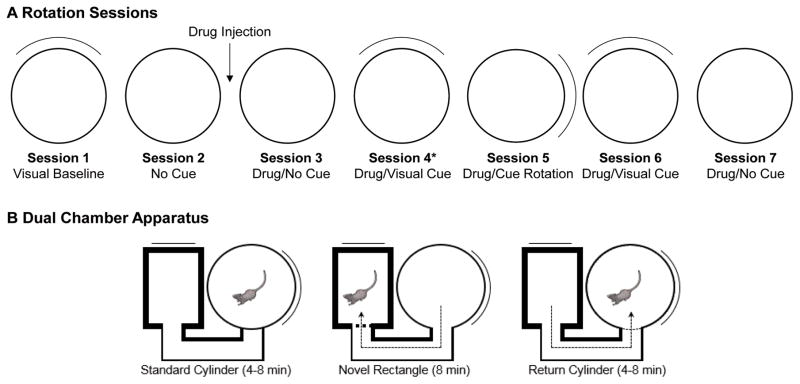
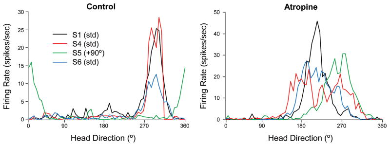
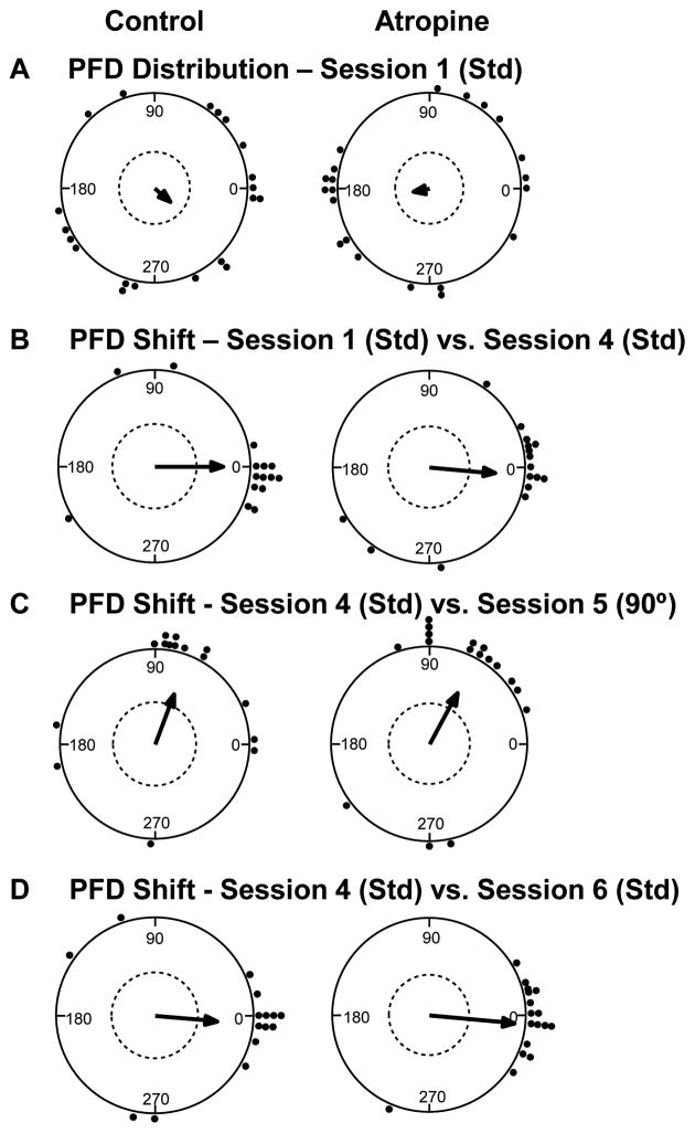
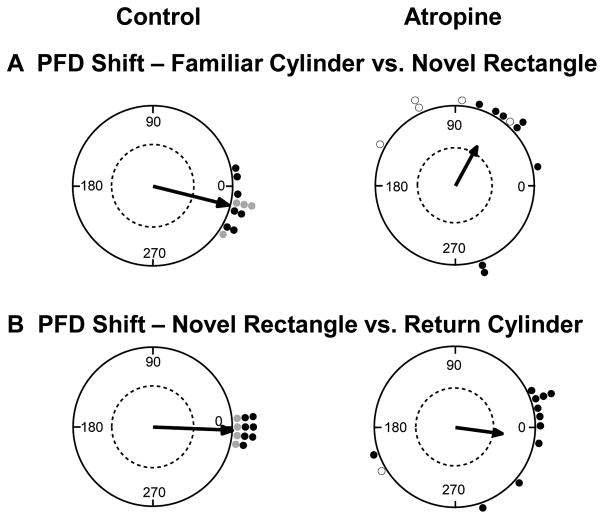
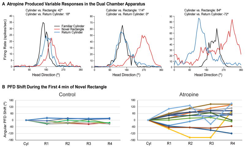
Acetylcholine contributes to accurate performance on some navigational tasks, but details of its contribution to the underlying brain signals are not fully understood. The medial septal area provides widespread cholinergic input to various brain regions, but selective damage to medial septal cholinergic neurons generally has little effect on landmark-based navigation, or the underlying neural representations of location and directional heading in visual environments. In contrast, the loss of medial septal cholinergic neurons disrupts navigation based on path integration, but no studies have tested whether these path integration deficits are associated with disrupted head direction (HD) cell activity. Therefore, we evaluated HD cell responses to visual cue rotations in a familiar arena, and during navigation between familiar and novel arenas, after muscarinic receptor blockade with systemic atropine. Atropine treatment reduced the peak firing rate of HD cells, but failed to significantly affect other HD cell firing properties. Atropine also failed to significantly disrupt the dominant landmark control of the HD signal, even though we used a procedure that challenged this landmark control. In contrast, atropine disrupted HD cell stability during navigation between familiar and novel arenas, where path integration normally maintains a consistent HD cell signal across arenas. These results suggest that acetylcholine contributes to path integration, in part, by facilitating the use of idiothetic cues to maintain a consistent representation of directional heading. (PsycINFO Database Record


Figures

Figure 1
Figure 1
Recording procedures used with the cylinder and dual-chamber apparatus. A, The animal was removed from the cylinder and placed in an opaque holding chamber following the first recording session. The floor paper was then changed, the cue card removed, and the animal was disoriented prior to the second session. At the end of the second session, the animal received an IP injection of saline or atropine sulfate, and placed into the holding chamber for 15 min. The animal was then replaced in the cylinder without disorientation. Following session 3, the cue card was inserted while the animal continued to navigate within the cylinder. For all subsequent sessions, the animal was removed and placed into the holding chamber, the floor paper was changed, and the animal was disoriented before the next session.
Figure 2
Figure 2
Representative tuning curves for HD cells in control (left) and atropine (right) conditions. For HD cells in both groups, the preferred direction remained consistent across standard recording sessions and shifted in the direction of the cue card during the rotation session. However, atropine treatment resulted in a significantly decreased peak firing rate and somewhat broader tuning curves, although this increased directional firing range was not significant.
Figure 3
Figure 3
HD cell response to cue card manipulations in the cylinder. A) The preferred directions of HD cells in both control (left) and atropine (right) groups initially showed a random distribution during the baseline recording session. B) The preferred direction remained relatively stable and well aligned with the cue card between standard (Std) sessions for both control and atropine groups. C) 90° rotation of the cue card resulted in a similar shift of the preferred direction for HD cells in both groups. D) The preferred firing direction returned to the original alignment for both groups, after the cue card was returned to its original position during the subsequent standard recording session.
Figure 4
Figure 4
Preferred firing direction (PFD) shifts in the dual chamber apparatus. A) Control HD cells (left) showed a relatively consistent preferred direction as the animal walked from the cylinder to the novel rectangle, suggesting the HD signal was maintained by path integration. In contrast, most HD cells in the atropine group (right) shifted toward the visual cue card (90° CCW), suggesting path integration failed to maintain the HD signal between arenas, and that the cue card in the rectangle may have driven the shift in the cells’ PFDs. B) Upon return to the familiar cylinder, control HD cells (left) realigned to the cue card in the cylinder. Atropine HD cells (right) also realigned to the cue card, albeit with less precision than the control group. Dashed line indicates .05 significance criterion. Open circles represent HD cells that shifted > 90° within the recording session. For the control group, black points represent HD cells recorded from the ADN (Taube & Burton, 1995) and gray points represent HD cells recorded from the lateral mammillary nuclei (Yoder et al., 2015).
Figure 5
Figure 5
Atropine treatment produced variable effects on HD cell activity during performance in the dual chamber apparatus. A) Some cells were influenced by the visual cue card, and then shifted to an intermediate direction upon return to the cylinder (left); other cells were influenced by the cue card and then returned precisely to the original alignment upon return (center); other cells drifted or were otherwise unstable within the rectangle, resulting in broad tuning curves (right). B) Temporal dynamics of HD cell activity in the novel rectangle. Control HD cells (left) remained relatively stable across the cylinder (Cyl) and the first four min (R1–R4) of the novel rectangle. In contrast, the preferred direction of many cells in the atropine group (right) drifted during the first four min of the rectangle session, relative to the cylinder.

References

    1. Aitken P, Benoit A, Zheng Y, Philoxene B, Le Gall A, Denise P, … Smith PF. Hippocampal and striatal M1 -muscarinic acetylcholine receptors are down-regulated following bilateral vestibular loss in rats. Hippocampus. 2016;26:1509–1514. doi: 10.1002/hipo.22651. - DOI - PubMed
    1. Bassett JP, Tullman ML, Taube JS. Lesions of the tegmentomammillary circuit in the head direction system disrupt the head direction signal in the anterior thalamus. J Neurosci. 2007;27:7564–7577. - PMC - PubMed
    1. Baxter MG, Bucci DJ, Sobel TJ, Williams MJ, Gorman LK, Gallagher M. Intact spatial learning following lesions of basal forebrain cholinergic neurons. Neuroreport. 1996;7:1417–1420. - PubMed
    1. Baxter MG, Gallagher M. Intact spatial learning in both young and aged rats following selective removal of hippocampal cholinergic input. Behav Neurosci. 1996a;110:460–467. - PubMed
    1. Baxter MG, Gallagher M. Neurobiological substrates of behavioral decline: models and data analytic strategies for individual differences in aging. Neurobiol Aging. 1996b;17:491–495. - PubMed