. 2017 Jul 17;12(7):e0180517.
doi: 10.1371/journal.pone.0180517. eCollection 2017.

Tales of diversity: Genomic and morphological characteristics of forty-six Arthrobacter phages

Karen K Klyczek  1 J Alfred Bonilla  1 Deborah Jacobs-Sera  2 Tamarah L Adair  3 Patricia Afram  4 Katherine G Allen  5 Megan L Archambault  1 Rahat M Aziz  6 Filippa G Bagnasco  3 Sarah L Ball  7 Natalie A Barrett  8 Robert C Benjamin  6 Christopher J Blasi  9 Katherine Borst  10 Mary A Braun  10 Haley Broomell  4 Conner B Brown  5 Zachary S Brynell  3 Ashley B Bue  1 Sydney O Burke  3 William Casazza  10 Julia A Cautela  8 Kevin Chen  10 Nitish S Chimalakonda  3 Dylan Chudoff  4 Jade A Connor  3 Trevor S Cross  4 Kyra N Curtis  3 Jessica A Dahlke  1 Bethany M Deaton  3 Sarah J Degroote  1 Danielle M DeNigris  8 Katherine C DeRuff  9 Milan Dolan  5 David Dunbar  4 Marisa S Egan  8 Daniel R Evans  10 Abby K Fahnestock  3 Amal Farooq  6 Garrett Finn  1 Christopher R Fratus  3 Bobby L Gaffney  5 Rebecca A Garlena  2 Kelly E Garrigan  8 Bryan C Gibbon  3 Michael A Goedde  5 Carlos A Guerrero Bustamante  2 Melinda Harrison  4 Megan C Hartwell  8 Emily L Heckman  11 Jennifer Huang  10 Lee E Hughes  6 Kathryn M Hyduchak  8 Aswathi E Jacob  8 Machika Kaku  10 Allen W Karstens  3 Margaret A Kenna  11 Susheel Khetarpal  10 Rodney A King  5 Amanda L Kobokovich  9 Hannah Kolev  10 Sai A Konde  3 Elizabeth Kriese  1 Morgan E Lamey  8 Carter N Lantz  3 Jonathan S Lapin  2 Temiloluwa O Lawson  1 In Young Lee  2 Scott M Lee  3 Julia Y Lee-Soety  8 Emily M Lehmann  1 Shawn C London  8 A Javier Lopez  10 Kelly C Lynch  5 Catherine M Mageeney  11 Tetyana Martynyuk  8 Kevin J Mathew  6 Travis N Mavrich  2 Christopher M McDaniel  5 Hannah McDonald  10 C Joel McManus  10 Jessica E Medrano  9 Francis E Mele  8 Jennifer E Menninger  8 Sierra N Miller  3 Josephine E Minick  3 Courtney T Nabua  8 Caroline K Napoli  8 Martha Nkangabwa  10 Elizabeth A Oates  5 Cassandra T Ott  2 Sarah K Pellerino  1 William J Pinamont  9 Ross T Pirnie  9 Marie C Pizzorno  9 Emilee J Plautz  1 Welkin H Pope  2 Katelyn M Pruett  3 Gabbi Rickstrew  10 Patrick A Rimple  2 Claire A Rinehart  5 Kayla M Robinson  6 Victoria A Rose  3 Daniel A Russell  2 Amelia M Schick  3 Julia Schlossman  10 Victoria M Schneider  2 Chloe A Sells  3 Jeremy W Sieker  3 Morgan P Silva  6 Marissa M Silvi  9 Stephanie E Simon  6 Amanda K Staples  5 Isabelle L Steed  1 Emily L Stowe  9 Noah A Stueven  1 Porter T Swartz  1 Emma A Sweet  1 Abigail T Sweetman  8 Corrina Tender  10 Katrina Terry  4 Chrystal Thomas  10 Daniel S Thomas  3 Allison R Thompson  5 Lorianna Vanderveen  10 Rohan Varma  10 Hannah L Vaught  1 Quynh D Vo  6 Zachary T Vonberg  1 Vassie C Ware  11 Yasmene M Warrad  3 Kaitlyn E Wathen  5 Jonathan L Weinstein  8 Jacqueline F Wyper  3 Jakob R Yankauskas  9 Christine Zhang  10 Graham F Hatfull  2
Affiliations

Tales of diversity: Genomic and morphological characteristics of forty-six Arthrobacter phages

Karen K Klyczek et al. PLoS One. .

Abstract

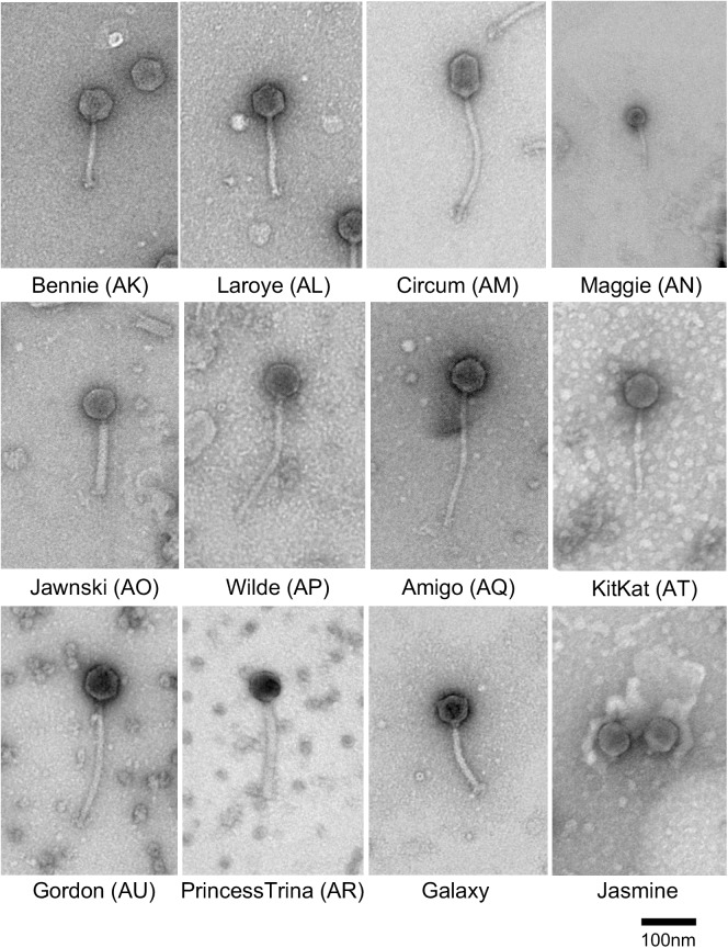
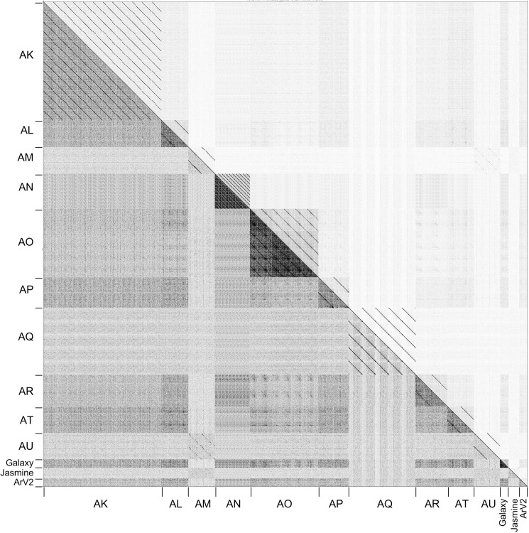
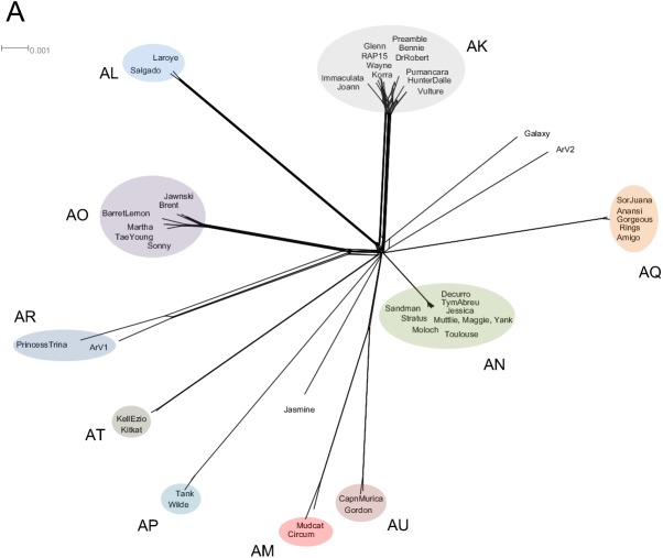
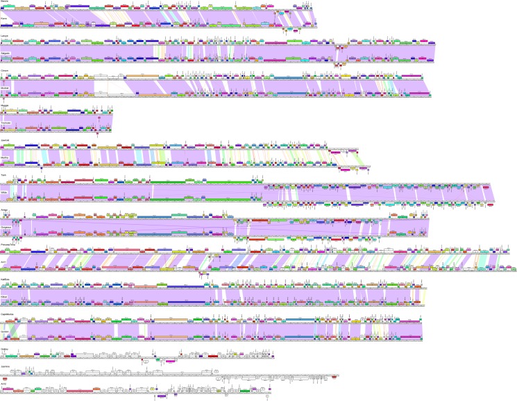
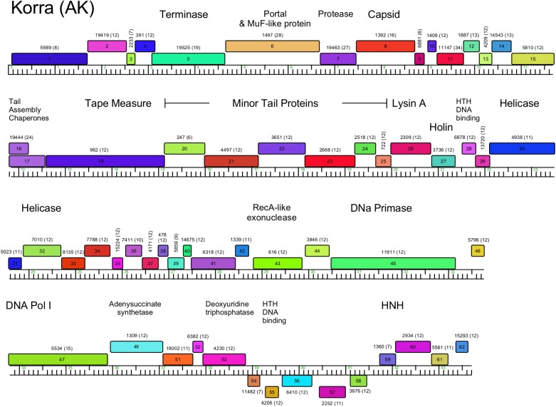
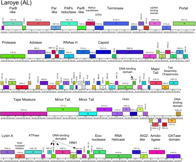
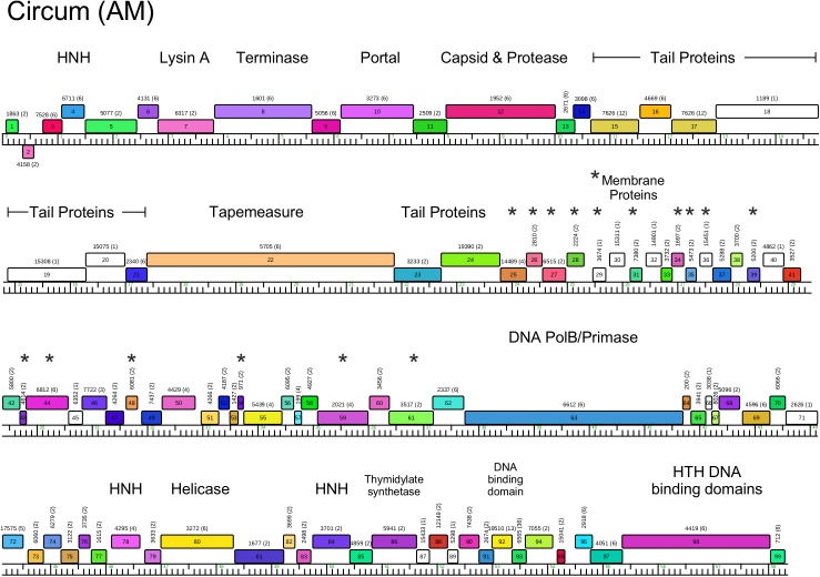
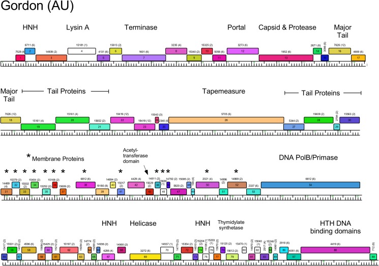
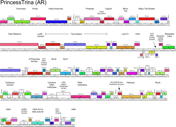
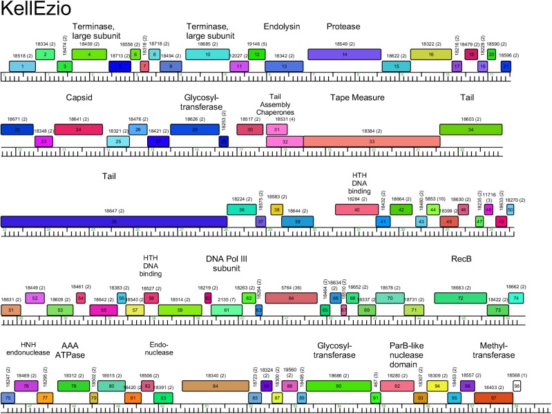
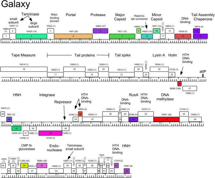
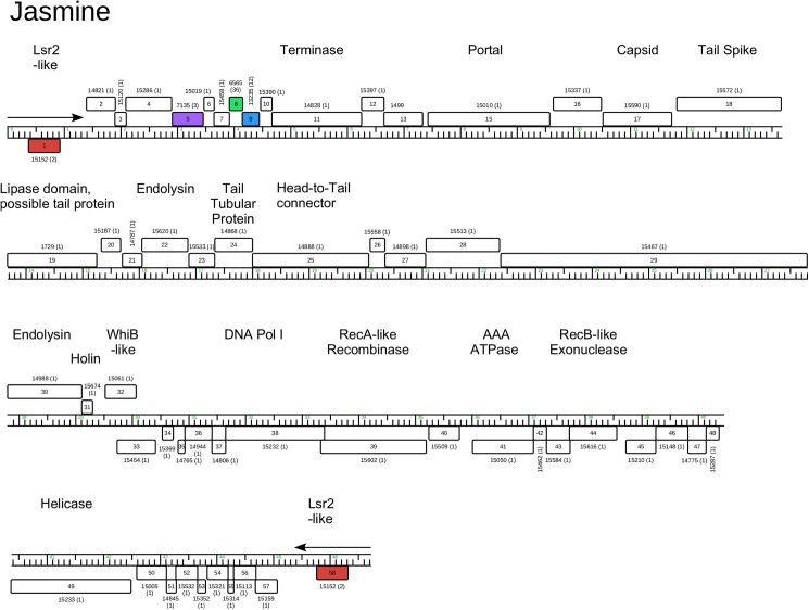
The vast bacteriophage population harbors an immense reservoir of genetic information. Almost 2000 phage genomes have been sequenced from phages infecting hosts in the phylum Actinobacteria, and analysis of these genomes reveals substantial diversity, pervasive mosaicism, and novel mechanisms for phage replication and lysogeny. Here, we describe the isolation and genomic characterization of 46 phages from environmental samples at various geographic locations in the U.S. infecting a single Arthrobacter sp. strain. These phages include representatives of all three virion morphologies, and Jasmine is the first sequenced podovirus of an actinobacterial host. The phages also span considerable sequence diversity, and can be grouped into 10 clusters according to their nucleotide diversity, and two singletons each with no close relatives. However, the clusters/singletons appear to be genomically well separated from each other, and relatively few genes are shared between clusters. Genome size varies from among the smallest of siphoviral phages (15,319 bp) to over 70 kbp, and G+C contents range from 45-68%, compared to 63.4% for the host genome. Although temperate phages are common among other actinobacterial hosts, these Arthrobacter phages are primarily lytic, and only the singleton Galaxy is likely temperate.


Conflict of interest statement

Competing Interests: The authors have declared that no competing interests exist.

Figures

Fig 1
Fig 1. Arthrobacter virion morphologies.
Electron micrographs of representative Arthrobacter phages. Scale bar corresponds to 100 nm.
Fig 2
Fig 2. Nucleotide sequence comparison of Arthrobacter phages.
Dot Plot of Arthrobacter phage genomes displayed using Gepard [35]. Individual genome sequences were concatenated into a single file arranged such that related genomes were adjacent to each other. The assignment of clusters is shown along both the left and bottom.
Fig 3
Fig 3. Splitstree representation of Arthrobacter phages and average nucleotide comparisons of Cluster AO Arthrobacter phages.
All Arthrobacter phage predicted proteins were assorted into 1052 phams according to shared amino acid sequence similarities. Each genome was then assigned a value reflecting the presence or absence of a pham member, and the genomes were compared and displayed using Splitstree [36]. Cluster and subcluster assignments derived from the dot plot and ANI analyses are annotated. The scale bar indicates 0.001 substitutions/site.
Fig 4
Fig 4. Pairwise alignment of clustered Arthrobacter phages.
The genomes of 23 Arthrobacter phages are shown. Pairwise nucleotide sequence similarity is displayed by color-spectrum coloring between the genomes, with violet as most similar and red as least similar. Genes are shown as boxes above (transcribed rightwards) and below (transcribed leftwards) each genome line; boxes are colored according to the gene phamilies they are assigned [29]. Maps were generated using Phamerator and its database Actinobacteriophage_692.
Fig 5
Fig 5. Genome organization of Arthrobacter phage Korra, Cluster AK.
The genome of Arthrobacter phage Korra is shown with predicted genes depicted as boxes either above (rightwards-expressed) or below (leftwards-expressed) the genome. Genes are colored according to the phamily designations using Phamerator and database Actinobacteriophage_692, with the phamily number shown above each gene with the number of phamily members in parentheses.
Fig 6
Fig 6. Genome organization of Arthrobacter phage Laroye, Cluster AL.
See Fig 5 for details.
Fig 7
Fig 7. Genome organization of Arthrobacter phage Circum, Cluster AM.
See Fig 5 for details.
Fig 8
Fig 8. Genome organization of Arthrobacter phage Gordon, Cluster AU.
See Fig 5 for details.
Fig 9
Fig 9. Genome organization of Arthrobacter phage Maggie, Cluster AN.
See Fig 5 for details.
Fig 10
Fig 10. Genome organization of Arthrobacter phage Jawnski, Cluster AO.
See Fig 5 for details.
Fig 11
Fig 11. Genome organization of Arthrobacter phage Tank, Cluster AP.
See Fig 5 for details.
Fig 12
Fig 12. Genome organization of Arthrobacter phage Amigo, Cluster AQ.
See Fig 5 for details.
Fig 13
Fig 13. Genome organization of Arthrobacter phage PrincessTrina, Cluster AR.
See Fig 5 for details.
Fig 14
Fig 14. Genome organization of Arthrobacter phage KellEzio, Cluster AT.
See Fig 5 for details.
Fig 15
Fig 15. Genome organization of Arthrobacter phage Galaxy, Singleton.
See Fig 5 for details.
Fig 16
Fig 16. Genome organization of Arthrobacter phage Jasmine, Singleton.
See Fig 5 for details.
Fig 17
Fig 17. Cluster diversity and inter-cluster relationships.
Intra-cluster diversity was determined by the percent of cluster-identifier phams (phams present in all members of a cluster and not found in phages of other clusters, red bars, not calculated for singleton phages), and the percent of orphams (phams present in only one phage, with no homologues in the database, blue bars). Inter-cluster relationships are shown as the proportion of phams present in each Arthrobacter phage cluster that are also present in at least one phage of another Arthrobacter cluster (yellow bars) or in at least one phage infecting a host other than Arthrobacter (green bars). The number of phages in each cluster is indicated in parentheses below the cluster name.
Fig 18
Fig 18. Comparison of phage shared gene content and host phylogeny.
A. One representative phage genome from each cluster including singletons were assigned a value reflecting the presence or absence of each pham in the database, and the genomes were compared and displayed using Splitstree [36]. Clusters are labeled with the cluster name, and singleton phages isolated in Arthrobacter are identified; all others are singleton phages isolated in other hosts. Colors correspond to bacterial host genera in panel B. The scale bar indicates 0.001 substitutions/site. B. Phylogenetic tree derived from 16S rRNA sequences from representative bacteria from each phage host genus in the database. Evolutionary analyses were conducted in MEGA7 [46] using the Neighbor-Joining method with gaps eliminated. The scale bar indicates 0.01 base substitutions per site. The 16S rRNA sequences (GenBank accession numbers in parentheses) were from Actinoplanes sp. SE50/110 (CP003170), Arthrobacter sp. ATCC 21022 (CP014196), Clavibacter michiganensis (AB299158), Corynebacterium vitaeruminis DSM 20294 (NR_121721), Gordonia terrae 3612 (CP016594), Microbacterium foliorum strain 122 (CP019892), M. smegmatis mc2 155 (Y08453), Propionibacterium acnes ATCC 11828 (CP003084), Rhodococcus erythropolis PR4 (AP008957), Streptomyces griseus strain DSM 40236 (AP009493), Tetraspheara remsis strain 3-M5-R-4 (DQ447774), Tsukamurella paurometabola DSM 20162 (NR_074458). This tree mirrors the phylogeny of 90 actinobacteria based on 16S rRNA gene sequences as described previously [47] but also includes Actinoplanes and Tetraspheara.

References

    1. Hendrix RW. Bacteriophages: evolution of the majority. Theor Popul Biol. 2002;61(4):471–80. . - PubMed
    1. Benson DA, Clark K, Karsch-Mizrachi I, Lipman DJ, Ostell J, Sayers EW. GenBank. Nucleic Acids Res. 2014;42(Database issue):D32–7. doi: 10.1093/nar/gkt1030 ; PubMed Central PMCID: PMCPMC3965104. - DOI - PMC - PubMed
    1. Dyson ZA, Tucci J, Seviour RJ, Petrovski S. Lysis to Kill: Evaluation of the Lytic Abilities, and Genomics of Nine Bacteriophages Infective for Gordonia spp. and Their Potential Use in Activated Sludge Foam Biocontrol. PLoS One. 2015;10(8):e0134512 doi: 10.1371/journal.pone.0134512 ; PubMed Central PMCID: PMCPMC4524720. - DOI - PMC - PubMed
    1. Liu M, Gill JJ, Young R, Summer EJ. Bacteriophages of wastewater foaming-associated filamentous Gordonia reduce host levels in raw activated sludge. Sci Rep. 2015;5:13754 doi: 10.1038/srep13754 ; PubMed Central PMCID: PMCPMC4563357. - DOI - PMC - PubMed
    1. Khairnar K, Pal P, Chandekar RH, Paunikar WN. Isolation and characterization of bacteriophages infecting nocardioforms in wastewater treatment plant. Biotechnol Res Int. 2014;2014:151952 doi: 10.1155/2014/151952 ; PubMed Central PMCID: PMCPMC4129933. - DOI - PMC - PubMed

LinkOut - more resources