. 2017 Aug 23;139(33):11434-11442.
doi: 10.1021/jacs.7b03966. Epub 2017 Aug 10.

Sulfur K-Edge XAS Studies of the Effect of DNA Binding on the [Fe4S4] Site in EndoIII and MutY

Affiliations

Sulfur K-Edge XAS Studies of the Effect of DNA Binding on the [Fe4S4] Site in EndoIII and MutY

Yang Ha et al. J Am Chem Soc. .

Abstract

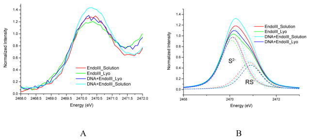
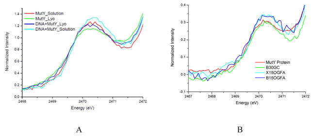
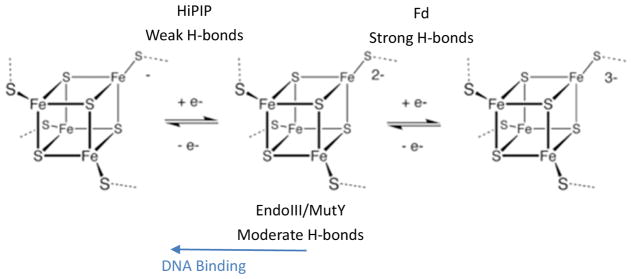
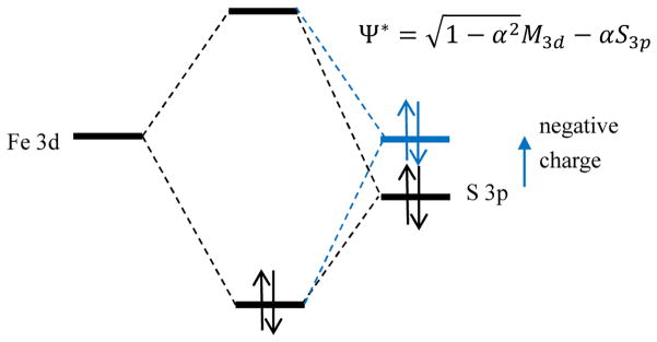
S K-edge X-ray absorption spectroscopy (XAS) was used to study the [Fe4S4] clusters in the DNA repair glycosylases EndoIII and MutY to evaluate the effects of DNA binding and solvation on Fe-S bond covalencies (i.e., the amount of S 3p character mixed into the Fe 3d valence orbitals). Increased covalencies in both iron-thiolate and iron-sulfide bonds would stabilize the oxidized state of the [Fe4S4] clusters. The results are compared to those on previously studied [Fe4S4] model complexes, ferredoxin (Fd), and to new data on high-potential iron-sulfur protein (HiPIP). A limited decrease in covalency is observed upon removal of solvent water from EndoIII and MutY, opposite to the significant increase observed for Fd, where the [Fe4S4] cluster is solvent exposed. Importantly, in EndoIII and MutY, a large increase in covalency is observed upon DNA binding, which is due to the effect of its negative charge on the iron-sulfur bonds. In EndoIII, this change in covalency can be quantified and makes a significant contribution to the observed decrease in reduction potential found experimentally in DNA repair proteins, enabling their HiPIP-like redox behavior.


Figures

Figure 1
Figure 1
S K-edge XAS of EndoIII without and with DNA and upon lyophilization (A) and the fits of the pre-edge region (B) using two peaks, one for the sulfides at ~2470.1 eV and one for the thiolates at ~2470.9 eV.
Figure 2
Figure 2
S K-edge XAS of MutY without and with non-specific DNA and upon lyophilization (A), and S-K edge spectra showing impact of different DNA length (15 or 30) and of specific (OGFA) as well as non-specific (GC) binding on MutY (B)
Figure 3
Figure 3
(A) Overlay of G. stearothermophilus (Gs) EndoIII (magenta, PDB 1ORN) and Gs MutY (cyan, PDB 5DPK), with [Fe4S4] cluster (orange for S and yellow for Fe atoms) and key Arg residues highlighted (red and blue respectively). Bound DNA in both structures are in green. Comparison of Ec EndoIII without DNA (pink, PDB 2ABK) in (B) and Gs EndoIII (magenta, PDB 1ORN) bound to DNA (green) in (C) showing the molecular surroundings of the [Fe4S4] cluster, including critical Arg residues (red). This comparison highlights the overall structure similarity between Gs EndoIII in complex with DNA to that of the Ec homolog without DNA. Small structural differences (RMSD100=1.4 Å) between the two homologs are attributed to their 43% sequence similarity.
Figure 3
Figure 3
(A) Overlay of G. stearothermophilus (Gs) EndoIII (magenta, PDB 1ORN) and Gs MutY (cyan, PDB 5DPK), with [Fe4S4] cluster (orange for S and yellow for Fe atoms) and key Arg residues highlighted (red and blue respectively). Bound DNA in both structures are in green. Comparison of Ec EndoIII without DNA (pink, PDB 2ABK) in (B) and Gs EndoIII (magenta, PDB 1ORN) bound to DNA (green) in (C) showing the molecular surroundings of the [Fe4S4] cluster, including critical Arg residues (red). This comparison highlights the overall structure similarity between Gs EndoIII in complex with DNA to that of the Ec homolog without DNA. Small structural differences (RMSD100=1.4 Å) between the two homologs are attributed to their 43% sequence similarity.
Figure 4
Figure 4
Local H-bond network of the Fe4S4 cluster for (A) HiPIP (PDB code: 1CKU), (B) EndoIII without DNA bound (PDB code: 4UNF), and EndoIII with DNA (PDB code: 1ORN) illustrating some of the H-bonds that could be affected by solvent water. The H-bonds are shown in dashed green, and the Arg and His are labeled. Schemes of the boxed region are shown in (D). In (A), an amide backbone H-bond to the thiolate S bound to Fe and an accessible solvent is shown in the box, and in (B) and (C), an Arg residue H-bond to the thiolate bound to Fe and accessible to solvent is shown in the box.
Figure 4
Figure 4
Local H-bond network of the Fe4S4 cluster for (A) HiPIP (PDB code: 1CKU), (B) EndoIII without DNA bound (PDB code: 4UNF), and EndoIII with DNA (PDB code: 1ORN) illustrating some of the H-bonds that could be affected by solvent water. The H-bonds are shown in dashed green, and the Arg and His are labeled. Schemes of the boxed region are shown in (D). In (A), an amide backbone H-bond to the thiolate S bound to Fe and an accessible solvent is shown in the box, and in (B) and (C), an Arg residue H-bond to the thiolate bound to Fe and accessible to solvent is shown in the box.
Scheme 1
Scheme 1
Fds have strong H-bonds to S (5 amide-thiolate H-bonds, 3 amide-sulfide H-bonds, as shown in Figure S6, and more importantly, H-bonds to surface exposed thiolate from solvent water), and thus both type of S donate less electron density to Fe, stabilizing the reduced state. (Redox potential range −700 to −300 mV) HiPIPs have only weak H-bonds to S (5 amide-thiolate H-bonds, as shown in Figure 4A), thus these S donate more electron density to Fe, stabilizing the oxidized state. (Redox potential range 100 to 400 mV) EndoIII/MutY have moderate H-bonding (A few amide-thiolate H-bonds, and importantly, an Arg-thiolate H-bond, as shown in Figure 4 and Figure S6), thus are redox inert. However, binding to DNA introduces negative charge, thus stabilizing the oxidized state.
Scheme 2
Scheme 2
Adding negative charges such as DNA in the proximity of the [Fe4S4] cluster destabilizes the S 3p orbital energy and increases the S 3p character (α2) in Ψ*. This increase in the Fe-S covalency stabilizes the oxidized more than the reduced state of the cluster and decreases the reduction potential.

References

    1. Holm RH, Kennepohl P, Solomon EI. Chem Rev. 1996;96:2239–2314. - PubMed
    1. Stiefel EI, George GN. Ferredoxins, Hydrogenases, and Nitrogenases: Metal-Sulfide Proteins. In: Bertini I, editor. Bioinorganic Chemistry. University Science Books; Mill Valley: 1994. pp. 365–453.
    1. Backes G, Mino Y, Loehr TM, Meyer TE, Cusanovich MA, Sweeney WV, Adman ET, Sanders-Loehr J. J Am Chem Soc. 1991;113:2055–2064.
    1. Teo BK, Shulman RG, Brown GS, Meixner AE. J Am Chem Soc. 1979;101:5624.
    1. Czernuszewicz RS, Macor KA, Johnson MK, Gewirth A, Spiro TG. J Am Chem Soc. 1987;109:7178–7187.

Publication types

MeSH terms