

PTSD Psychotherapy Outcome Predicted by Brain Activation During Emotional Reactivity and Regulation
- PMID: 28715908
- PMCID: PMC5711543
- DOI: 10.1176/appi.ajp.2017.16091072
PTSD Psychotherapy Outcome Predicted by Brain Activation During Emotional Reactivity and Regulation
Abstract
Objective: Exposure therapy is an effective treatment for posttraumatic stress disorder (PTSD), but many patients do not respond. Brain functions governing treatment outcome are not well characterized. The authors examined brain systems relevant to emotional reactivity and regulation, constructs that are thought to be central to PTSD and exposure therapy effects, to identify the functional traits of individuals most likely to benefit from treatment.
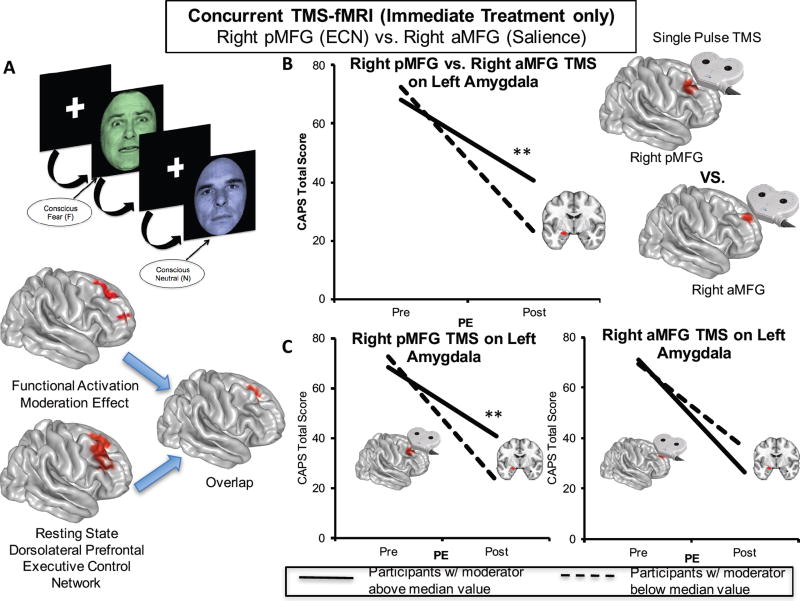
Method: Individuals with PTSD underwent functional MRI (fMRI) while completing three tasks assessing emotional reactivity and regulation. Participants were then randomly assigned to immediate prolonged exposure treatment (N=36) or a waiting list condition (N=30). A random subset of the prolonged exposure group (N=17) underwent single-pulse transcranial magnetic stimulation (TMS) concurrent with fMRI to examine whether predictive activation patterns reflect causal influence within circuits. Linear mixed-effects modeling in line with the intent-to-treat principle was used to examine how baseline brain function moderated the effect of treatment on PTSD symptoms.
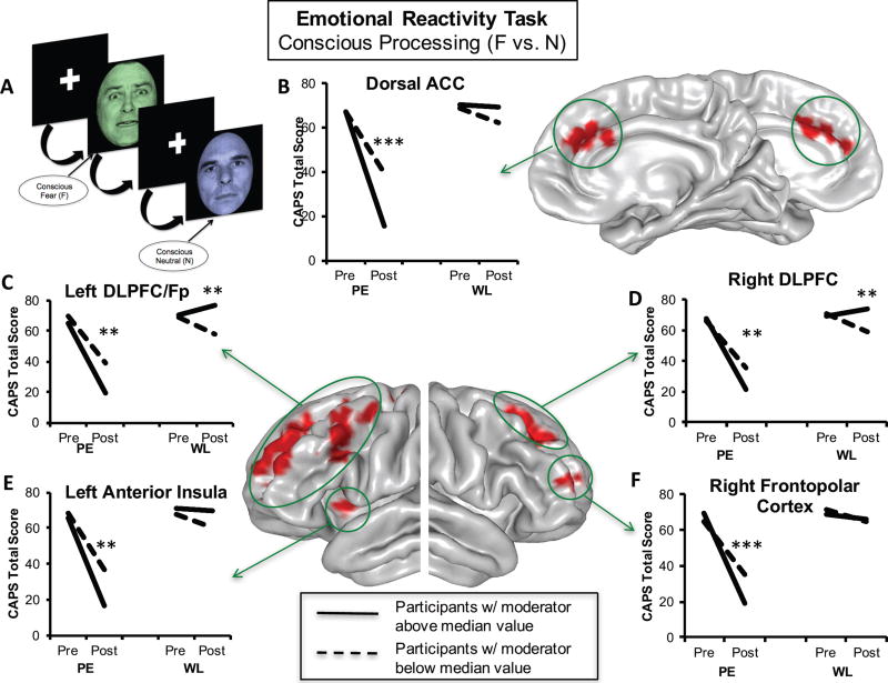
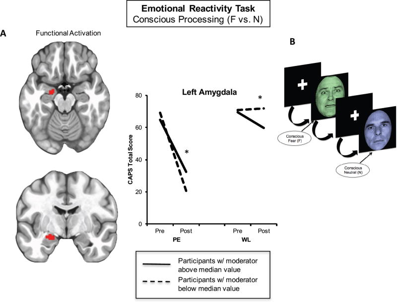
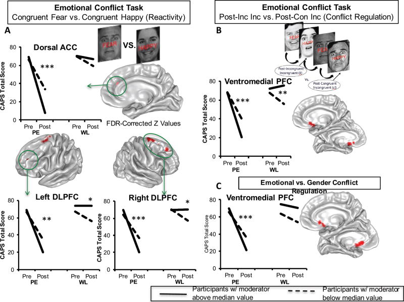
Results: At baseline, individuals with larger treatment-related symptom reductions (compared with the waiting list condition) demonstrated 1) greater dorsal prefrontal activation and 2) less left amygdala activation, both during emotion reactivity; 3) better inhibition of the left amygdala induced by single TMS pulses to the right dorsolateral prefrontal cortex; and 4) greater ventromedial prefrontal/ventral striatal activation during emotional conflict regulation. Reappraisal-related activation was not a significant moderator of the treatment effect.
Conclusions: Capacity to benefit from prolonged exposure in PTSD is gated by the degree to which prefrontal resources are spontaneously engaged when superficially processing threat and adaptively mitigating emotional interference, but not when deliberately reducing negative emotionality.
Trial registration: ClinicalTrials.gov NCT01507948.
Keywords: Brain Imaging Techniques; Emotion; Exposure Therapy; Posttraumatic Stress Disorder; Psychotherapy; Transcranial Magnetic Stimulation.
Conflict of interest statement
Disclosures
All other authors report no financial conflicts of interest.
Figures
Comment in
-
Frontal Lobe Moderators and Mediators of Response to Exposure Therapy in PTSD.Am J Psychiatry. 2017 Dec 1;174(12):1131-1133. doi: 10.1176/appi.ajp.2017.17091056. Am J Psychiatry. 2017. PMID: 29191039 No abstract available.
Similar articles
-
Selective Effects of Psychotherapy on Frontopolar Cortical Function in PTSD.Am J Psychiatry. 2017 Dec 1;174(12):1175-1184. doi: 10.1176/appi.ajp.2017.16091073. Epub 2017 Jul 18. Am J Psychiatry. 2017. PMID: 28715907 Free PMC article. Clinical Trial.
-
Neural correlates of emotional reactivity and regulation associated with treatment response in a randomized clinical trial for posttraumatic stress disorder.Psychiatry Res Neuroimaging. 2020 May 30;299:111062. doi: 10.1016/j.pscychresns.2020.111062. Epub 2020 Mar 5. Psychiatry Res Neuroimaging. 2020. PMID: 32278278 Free PMC article. Clinical Trial.
-
History of childhood maltreatment augments dorsolateral prefrontal processing of emotional valence in PTSD.J Psychiatr Res. 2016 Mar;74:45-54. doi: 10.1016/j.jpsychires.2015.12.015. Epub 2015 Dec 19. J Psychiatr Res. 2016. PMID: 26741277 Free PMC article.
-
Magnetic resonance imaging predictors of psychotherapy treatment response in post-traumatic stress disorder: A role for the salience network.Psychiatry Res. 2019 Jul;277:52-57. doi: 10.1016/j.psychres.2019.02.005. Epub 2019 Feb 2. Psychiatry Res. 2019. PMID: 30755338 Review.
-
Amygdala, medial prefrontal cortex, and hippocampal function in PTSD.Ann N Y Acad Sci. 2006 Jul;1071:67-79. doi: 10.1196/annals.1364.007. Ann N Y Acad Sci. 2006. PMID: 16891563 Review.
Cited by
-
Associations between resting-state functional connectivity and treatment response in a randomized clinical trial for posttraumatic stress disorder.Depress Anxiety. 2020 Oct;37(10):1037-1046. doi: 10.1002/da.23075. Epub 2020 Jul 15. Depress Anxiety. 2020. PMID: 32668087 Free PMC article. Clinical Trial.
-
Individual prediction of psychotherapy outcome in posttraumatic stress disorder using neuroimaging data.Transl Psychiatry. 2019 Dec 2;9(1):326. doi: 10.1038/s41398-019-0663-7. Transl Psychiatry. 2019. PMID: 31792202 Free PMC article.
-
Predicting antidepressant responsiveness in major depressive disorder patients via electroencephalography gamma-band dynamic functional connectivity in response to salient auditory stimuli.Int J Neuropsychopharmacol. 2025 Jul 23;28(7):pyaf042. doi: 10.1093/ijnp/pyaf042. Int J Neuropsychopharmacol. 2025. PMID: 40577659 Free PMC article.
-
Case Series: Unilateral Amygdala Ablation Ameliorates Post-Traumatic Stress Disorder Symptoms and Biomarkers.Neurosurgery. 2020 Sep 15;87(4):796-802. doi: 10.1093/neuros/nyaa051. Neurosurgery. 2020. PMID: 32259241 Free PMC article.
-
Neural function during emotion processing and modulation associated with treatment response in a randomized clinical trial for posttraumatic stress disorder.Depress Anxiety. 2020 Jul;37(7):670-681. doi: 10.1002/da.23022. Epub 2020 Apr 19. Depress Anxiety. 2020. PMID: 32306485 Free PMC article. Clinical Trial.
References
-
- Foa EB, Gillihan SJ, Bryant RA. Challenges and Successes in Dissemination of Evidence-Based Treatments for Posttraumatic Stress: Lessons Learned From Prolonged Exposure Therapy for PTSD. Psychological science in the public interest : a journal of the American Psychological Society. 2013;14:65–111. - PMC - PubMed
-
- Bradley R, Greene J, Russ E, Dutra L, Westen D. A multidimensional meta-analysis of psychotherapy for PTSD. Am J Psychiatry. 2005;162:214–227. - PubMed
Publication types
MeSH terms
Associated data
Grants and funding
LinkOut - more resources
Full Text Sources
Other Literature Sources
Medical
