Review
. 2017 Sep 15:3:167-196.
doi: 10.1146/annurev-vision-102016-061214. Epub 2017 Jul 17.

The Functional Neuroanatomy of Human Face Perception

Affiliations
Review

The Functional Neuroanatomy of Human Face Perception

Kalanit Grill-Spector et al. Annu Rev Vis Sci. .

Abstract

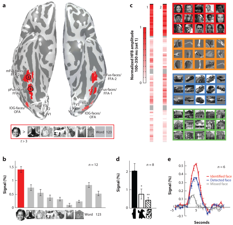
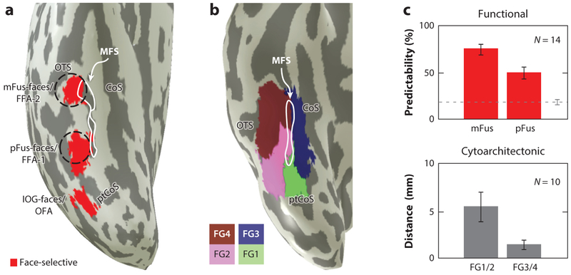
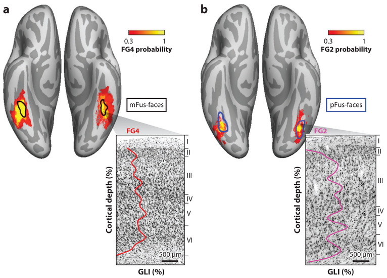
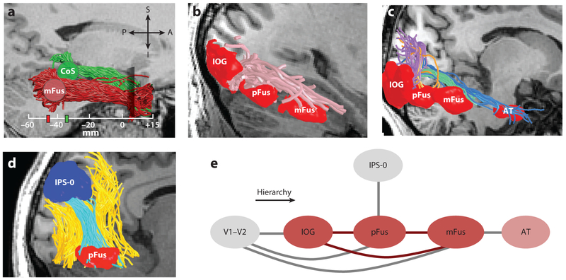
Face perception is critical for normal social functioning and is mediated by a network of regions in the ventral visual stream. In this review, we describe recent neuroimaging findings regarding the macro- and microscopic anatomical features of the ventral face network, the characteristics of white matter connections, and basic computations performed by population receptive fields within face-selective regions composing this network. We emphasize the importance of the neural tissue properties and white matter connections of each region, as these anatomical properties may be tightly linked to the functional characteristics of the ventral face network. We end by considering how empirical investigations of the neural architecture of the face network may inform the development of computational models and shed light on how computations in the face network enable efficient face perception.

Keywords: FFA; fMRI; face network; face recognition; mid-fusiform sulcus; population receptive fields; ventral stream.


Figures

Figure 1
Figure 1
Face-selective regions in human ventral occipito-temporal cortex. (a) Face-selective regions are identified on the basis of higher responses to faces compared to a variety of other stimuli (faces > bodies, objects, places, and characters; t > 3, voxel level). As concluded in our prior work (Weiner & Grill-Spector 2010, 2011, 2012, 2013), including many categories in the statistical contrast (as opposed to faces > objects or faces > places) improves the accuracy of delineating face-selective regions in ventral temporal cortex (VTC). Additionally, localizing body-selective regions further guides the parcellation, as body- and face-selective regions together form a topographic map in VTC (Orlov et al. 2010; Weiner & Grill-Spector 2010, 2011, 2012, 2013). The figure shows an inflated cortical surface of an individual participant depicting the typical three clusters of face-selective regions in ventral occipito-temporal cortex. These clusters are found bilaterally and arranged from posterior to anterior. One cluster is on the inferior occipital gyrus (IOG) referred to as IOG-faces [also as the occipital face area (OFA)]; a second cluster is on the posterior aspect of the fusiform gyrus, extending to the occipito-temporal sulcus, referred to as pFus-faces [also fusiform face area one (FFA-1)]; a third cluster is located approximately 1–1.5 cm more anterior on the lateral aspect of the fusiform gyrus, overlapping the anterior tip of the mid-fusiform sulcus (MFS) and is referred to as mFus-faces (also referred to as FFA-2). White lines: boundaries of retinotopic areas. (b) Response amplitudes of mFus-faces from independent data showing the typical higher responses to faces compared to other stimuli. Adapted from Stigliani et al. (2015). (c) Responses to single images in pFus- and mFus-faces from electrocorticography recordings. Each cell shows the normalized responses in the high frequency broadband range (HFB) (30–150 Hz) to a single image averaged across 2–5 presentations of that image over a 100–350-ms time window. The first column shows responses in an electrode over pFus-faces/FFA-1 (indicated by 1 in panel a), and the second shows responses over mFus-faces/FFA-2 (indicated by 2 in panel a). Responses to face images that vary in gender, expression, size, pose, and background are higher than any of the nonface images. Adapted from Jacques et al. (2016). (d) Responses in ventral face-selective regions to face silhouettes are significantly higher than two-tone shapes and scrambled images. Adapted from Davidenko et al. (2012). (e) Responses in ventral face-selective regions are highest when faces are identified, intermediate when they are detected but not identified, and lowest when they are missed. Adapted from Grill-Spector et al. (2004).
Figure 2
Figure 2
Face-selective regions and cytoarchitectonic boundaries are predicted by cortical folding. (a) Inflated cortical surface zoomed on ventral temporal cortex (VTC) of the right hemisphere of a representative participant showing face-selective regions (faces > other categories; t > 3, voxel level) in red. White: mid-fusiform sulcus (MFS). Dashed black lines: 1-cm diameter disks aligned to the anterior and posterior tips of the MFS, respectively. (b) Inflated cortical surface zoomed on VTC of the right hemisphere of a representative postmortem brain showing cytoarchitectonic areas within the fusiform gyrus (FG), each in a distinct color (see legend). White: MFS. (c, top) Percentage of face-selective voxels predicted by disks in panel a. Dashed black line: percentage of face-selective voxels predicted by Talairach coordinates. Error bars: standard error of the mean across participants. There is a tighter coupling between the MFS and mFus-faces/FFA-2 located more anteriorly than pFus-faces/FFA-1, which is located more posteriorly. Adapted from Weiner et al. (2014). (Bottom) The distance between the MFS and the boundary between cytoarchitectonic areas of the medial FG (FG1 or FG3) and the lateral FG (FG2 or FG4). There is a tighter coupling between the MFS and the anterior cytoarchitectonic boundary between FG3 and FG4 compared to the posterior boundary between FG1 and FG2. Adapted from Lorenz et al. (2017). Abbreviations: CoS, collateral sulcus; OFA, occipital face area; OTS, occipito-temporal sulcus; pFus, posterior fusiform; mFus, mid fusiform.
Figure 3
Figure 3
mFus- and pFus-faces are cytoarchitectonically dissociable. (a) mFus-faces/FFA-2 is located in cytoarchitectonic area FG4. (b) pFus-faces/FFA-1 is located in cytoarchitectonic area FG2. In each panel (top): superimposition of the probabilistic map of a cytoarchitectonic area (see colorbar) and the probabilistic definition of a functional area (contour) on the FreeSurfer average brain. In each panel (bottom): cell-body-stained histological section and the gray level index (GLI; line) across cortical layers from a representative 20-micron slice. Cytoarchitectonic maps used data from 10 postmortem brains. Functional definitions are based on data from 12 living subjects.
Figure 4
Figure 4
White matter tracts of the ventral face network. (a) Separate parallel tracts are associated with mFus-faces/FFA-2 (red) and CoS-places/PPA (green). Tracts were identified on the basis of functional regions and an anatomical plane in the anterior ventral temporal lobe used to define the inferior longitudinal fasciculus. (b) White matter tracts directly interconnecting IOG-, pFus-, and mFus-faces. (c) Connections from early visual retinotopic regions (V1–V2) to IOG-faces (purple), pFus-faces (orange), mFus-faces (green), and a face-selective region in anterior temporal cortex (AT; blue). (d) A subset of the vertical occipital fasciculus (yellow) connects IPS-0 and pFus-faces (cyan). Data in panels ad show representative data from individual participants. In each panel, the brain section is illustrated in gray and shown in a sagittal view. White matter tracts are illustrated as colored tubes. Face-selective regions illustrated in red were dilated from the gray matter to extend to the white matter. (e) A schematic summarizing results of tractography experiments. Dark red ovals: core face-selective regions; pink oval: non-core face-selective region; gray ovals: regions that are considered external to the face network. The schematic is arranged such that the hierarchical axis is from left to right. Abbreviations: A, anterior; AT, anterior temporal; CoS, collateral sulcus; I, inferior; IOG, inferior occipital gyrus; IPS, intraparietal sulcus; P, posterior; PPA, parahippocampal place area; S, superior.
Figure 5
Figure 5
Properties of functionally defined white matter correlate with face recognition. (a) Correlation between performance (% correct) in the Benton Facial Recognition Test (Benton 1980) and the fractional anisotropy (FA) of white matter next to mFus-faces [referred to as functionally defined white matter] in typical participants. Each point represents data from a single subject. Line: the line-of-best-fit to the data showing a positive correlation between behavior and FA; shaded area: 95% confidence interval. (b) Whole: mean diffusivity (MD) averaged across a longitudinal white matter tract connected to mFus-faces (see Figure 4a). Local: MD averaged in the local white matter adjacent to mFus-faces. Red: typical adults; gray: adults with developmental prosopagnosia (DP). Each point represents a participant. MD is lower in adults with DP than in typical controls. Asterisk: p < 0.01. Adapted from Gomez et al. (2015).
Figure 6
Figure 6
Population receptive fields (pRFs) reveal a hierarchical organization of the ventral face network. Population receptive fields were estimated from functional magnetic resonance imaging responses to faces presented at different visual field locations. For the data in this panel, pRFs were estimated in a task during which participants fixated on a central crosshair and reported when they detected a small dot superimposed on some of the faces. (a) Median eccentricity of pRF centers for areas constituting the ventral visual stream from V1 to mFus-faces. Error bars: 68% confidence intervals (CIs). (b) Median pRF size for each area from V1 to mFus-faces. Error bars: 68% CIs. Population-receptive-field size is defined as the standard deviation of a two-dimensional Gaussian that characterizes the response of the pRF to point stimuli. (c) Relationship between pRF size and eccentricity. Shaded area: 68% CI on a line fitted to the data. (d) Facial features processed by example pRFs across the ventral visual processing hierarchy. Circles: pRFs at 1° eccentricity (derived from panel c). Each circle is drawn at ± 2 pRF sizes. The depicted face is sized to simulate a face seen from a conversational distance of 1 m, approximately 6.5°, based on average male head sizes (Loftus & Harley 2005, McKone 2009). Ascending the hierarchy, spatial information is integrated across increasingly larger regions of the face until the latest stages where entire faces are processed by the neural population within a voxel. Adapted from Kay et al. (2015). Abbreviation: IOG, inferior occipital gyrus.
Figure 7
Figure 7
Attention modulates population receptive field (pRF) properties in the ventral face network, enhancing spatial representations. Population receptive fields were measured under different tasks using the same stimulus. For the data in this panel, subjects performed either a one-back task on centrally presented digits (digit task) or a one-back task on the presented face (face task). (a) Task-induced changes in pRF properties. Bars: median across voxels; error bars: 68% confidence intervals (CIs). pRFs in IOG-, pFus-, and mFus-faces, and hV4 are larger (left), are more eccentric (middle), and have increased gain (right) during the face task (blue) compared to the digit task (red). (b) Tiling of visual field by 100 randomly selected pRFs from left pFus-faces. Dots: pRF centers; circles: pRFs drawn at ± 2 pRF sizes; intensity: pRF gain (see colorbar). An example face is shown at 5° eccentricity. (c) Spatial uncertainty in discrimination of stimulus positions. Bars: amount of uncertainty for reference positions at 5° eccentricity (median across angular positions ± 68% CI). During the digit task, uncertainty in IOG-, pFus-, and mFus-faces is large. However, during the face task, uncertainty is substantially reduced and is commensurate with the uncertainty in V1. Adapted from Kay et al. (2015). Abbreviation: IOG, inferior occipital gyrus.

References

    1. Allison T, Ginter H, McCarthy G, Nobre AC, Puce A, et al. 1994a. Face recognition in human extrastriate cortex. J. Neurophysiol 71:821–25 - PubMed
    1. Allison T, McCarthy G, Nobre A, Puce A, Belger A. 1994b. Human extrastriate visual cortex and the perception of faces, words, numbers, and colors. Cereb. Cortex 4:544–54 - PubMed
    1. Allison T, Puce A, Spencer DD, McCarthy G. 1999. Electrophysiological studies of human face perception. I: potentials generated in occipitotemporal cortex by face and non-face stimuli. Cereb. Cortex 9:415–30 - PubMed
    1. Amunts K, Malikovic A, Mohlberg H, Schormann T, Zilles K. 2000. Brodmann’s areas 17 and 18 brought into stereotaxic space—where and how variable? NeuroImage 11:66–84 - PubMed
    1. Amunts K, Zilles K. 2015. Architectonic mapping of the human brain beyond Brodmann. Neuron 88:1086–107 - PubMed

Publication types

LinkOut - more resources