. 2017 Jul 17;16(1):124.
doi: 10.1186/s12943-017-0700-1.

FOXP3 promotes tumor growth and metastasis by activating Wnt/β-catenin signaling pathway and EMT in non-small cell lung cancer

Affiliations

FOXP3 promotes tumor growth and metastasis by activating Wnt/β-catenin signaling pathway and EMT in non-small cell lung cancer

Shucai Yang et al. Mol Cancer. .

Abstract

Background: The role of cancer cell FOXP3 in tumorigenesis is conflicting. We aimed to study FOXP3 expression and regulation, function and clinical implication in human non-small cell lung cancer (NSCLC).

Methods: One hundred and six patients with histologically-confirmed NSCLC who underwent surgery were recruited for the study. Tumor samples and NSCLC cell lines were used to examine FOXP3 and its related molecules. Various cell functions related to tumorigenesis were performed. In vivo mouse tumor xenograft was used to confirm the in vitro results.

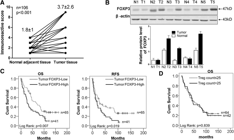
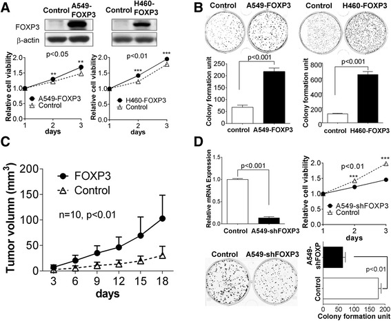
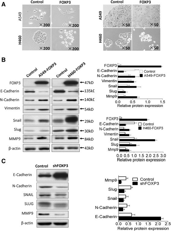
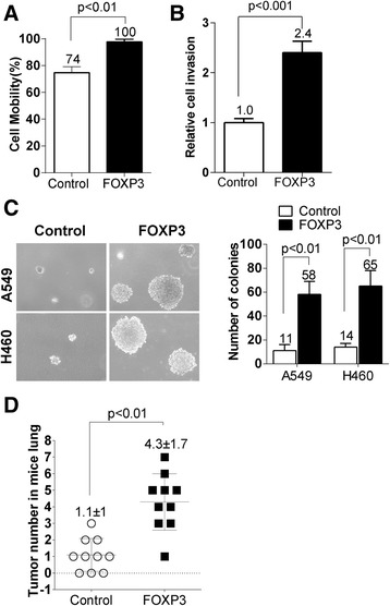
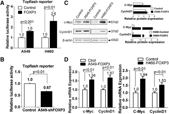
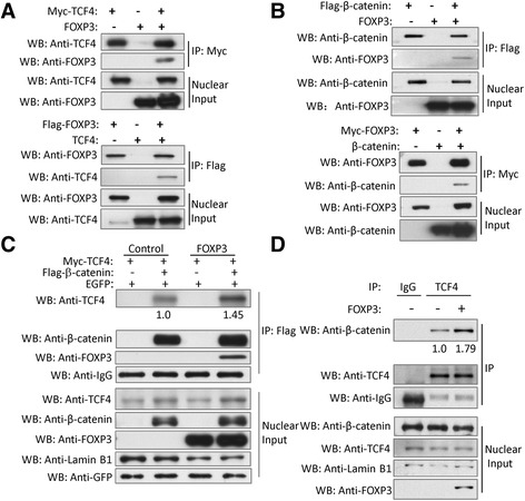
Results: NSCLC patients with the high level of FOXP3 had a significant decrease in overall survival and recurrence-free survival. FOXP3 overexpression significantly induced cell proliferation, migration, and invasion, whereas its inhibition impaired its oncogenic function. In vivo studies confirmed that FOXP3 promoted tumor growth and metastasis. The ectopic expression of FOXP3 induced epithelial-mesenchymal transition (EMT) with downregulation of E-cadherin and upregulation of N-cadherin, vimentin, snail, slug, and MMP9. The oncogenic effects by FOXP3 could be attributed to FOX3-mediated activation of Wnt/β-catenin signaling, as FOXP3 increased luciferase activity of Topflash reporter and upregulated Wnt signaling target genes including c-Myc and Cyclin D1 in NSCLC cells. Co-immunoprecipitation results further indicated that FOXP3 could physically interacted with β-catenin and TCF4 to enhance the functions of β-catenin and TCF4, inducing transcription of Wnt target genes to promote cell proliferation, invasion and EMT induction.

Conclusions: FOXP3 can act as a co-activator to facilitate the Wnt-b-catenin signaling pathway, inducing EMT and tumor growth and metastasis in NSCLC.

Keywords: EMT; FOXP3; NSCLC; TCF4; Wnt.


Conflict of interest statement

Ethics approval and consent to participate

Human studies were approved by the Joint CUHK-NTEC Clinical Research Ethics committee. All procedures of mouse experiments were approved by the Animal Ethics Committee of the Chinese University of Hong Kong.

Consent for publication

Not applicable.

Competing interests

The authors declare that they have no competing interests.

Publisher’s Note

Springer Nature remains neutral with regard to jurisdictional claims in published maps and institutional affiliations.

Figures

Fig. 1
Fig. 1
FOXP3 is highly expressed in NSCLC and correlated with poor prognosis. a The levels of FOXP3 in 106 paired NSCLC tissues and adjacent normal tissues were scored by IRS method (5) and analyzed by paired t-test. Mean ± SD is shown in the figure. For images of the tissue staining, please refer to Additional file 2: Figure S1. b FOXP3 protein levels were higher in tumor than in peritumoral tissues, as determined by western blot. c Kaplan–Meier representation of the overall survival and recurrence-free survival of the two groups of patients with high (n = 41, solid line) or low (n = 65, dotted line) FOXP3 expression in NSCLC tissues. Statistical analysis was performed with the log-rank test. d Kaplan–Meier representation of the overall survival of the two groups of patients with Treg cell counts >25 (n = 42, solid line) or ≤25 (n = 64, dotted line) in NSCLC tissues. Statistical analysis was performed with the log-rank test
Fig. 2
Fig. 2
FOXP3 promotes tumor growth in NSCLC. a Effect of FOXP3 on cell viability was evaluated by MTT assay in A549 and H460 cells. b Effect of FOXP3 on colony formation in A549 and H460 cells. c Subcutaneous tumor growth curve of A549-FOXP3 cells in nude mice was compared with control cells. The FOXP3 group showed an enhanced tumor growth compared with the control group (P < 0.05). d Effects of FOXP3 knockdown on cell viability and colony formation in A549 cells
Fig. 3
Fig. 3
FOXP3 induces EMT in NSCLC cells. a FOXP3 induces mesenchymal morphology changes in A549 and H460 cells: spindle shape and loss of cell-cell contact. b The effect of FOXP3 overexpression on EMT marker expression was assessed by western blot. c Effect of FOXP3 knockdown on EMT marker expression in A549 cells
Fig. 4
Fig. 4
FOXP3 promotes tumor metastasis in NSCLC. a Effect of FOXP3 on cell migration was evaluated by wound healing assay in A549 cells. FOXP3-expressing A549 cells had a much stronger healing ability than control cells (p < 0.01). b Effect of FOXP3 on cell invasion was evaluated in A549 cells by Matrigel invasion assay. FOXP3-expressing A549 cells showed higher penetration rate through the Matrigel-coated membrane compared with control cells (p < 0.001). c Soft agar growth analysis demonstrated that FOXP3-expressing A549 and H460 cells had a stronger tumorigenicity than control cells (p < 0.01). d FOXP3 promotes tumor metastasis in vivo. The total number of metastatic nodules was quantified in lungs of nude mice 8 weeks after tail vein injection of control and FOXP3-expressing A549 cells. Values for individual mouse were dotted in the plot and values by group were also denoted. Data are mean ± s.d. Please refer to Additional file 2: Figure S4 for representative images for a, b and d
Fig. 5
Fig. 5
The oncogenic effect of FOXP3 is mediated by activating Wnt/β-catenin signaling pathway. a The effect of FOXP3 on Wnt/β-catenin signaling pathway was assessed by dual-luciferase reporter assays in A549 and H460 cells. b Knockdown of FOXP3 impaired Topflash reporter activities in A549 cells. c FOXP3 upregulated protein expression of c-Myc and Cyclin D1 in A549 and H460 cells. d FOXP3 upregulated mRNA expression of c-Myc and Cyclin D1 in A549 and H460 cells
Fig. 6
Fig. 6
FOXP3 acts as a co-activator of Wnt/β-catenin signaling in NSCLC cells. a Co-immunoprecipitation showed the physical interaction between FOXP3 and TCF4 in HEK-293 cells co-expressing Myc-TCF4 and FOXP3 or Flag-FOXP3 and TCF4. b Co-immunoprecipitation showed the physical interaction between FOXP3 and β-catenin in HEK-293 cells co-expressing Flag-β-catenin and FOXP3 or Myc-FOXP3 and β-catenin. c FOXP3 enhances the interaction of β-catenin and TCF4. Plasmids of Myc-TCF4, Flag-β-catenin and FOXP3 were co-transfected into HEK293T cells according to the situations indicated above. EGFP plasmids was co-transfected as the control of transfection efficiency. The cell lysates were subjected to IP with an anti-Flag antibody. The immunoprecipitates and the nuclear proteins were subjected to Western blots using the indicated antibodies. Lamin B1 was used to be the loading control of the nuclear protein. d FOXP3 enhances the interaction of endogenous TCF4 and β-catenin. A549 cells were infected with FOXP3 lentivirus or control lentivirus as indicated above for 48 h. The nuclear fractions were incubated with an anti-TCF4 antibody for the IP experiment. IgG was used as a negative control. The immunoprecipitates and the nuclear extracts were subjected to Western blots using the indicated antibodies. Lamin B1 was used to be the loading control of the nuclear protein

References

    1. Hinz S, Pagerols-Raluy L, Oberg HH, Ammerpohl O, Grussel S, Sipos B, Grützmann R, Pilarsky C, Ungefroren H, Saeger HD, et al. Foxp3 expression in pancreatic carcinoma cells as a novel mechanism of immune evasion in cancer. Cancer Res. 2007;67:8344–8350. doi: 10.1158/0008-5472.CAN-06-3304. - DOI - PubMed
    1. Liang YJ, Liu HC, Su YX, Zhang TH, Chu M, Liang LZ, Liao GQ. Foxp3 expressed by tongue squamous cell carcinoma cells correlates with clinicopathologic features and overall survival in tongue squamous cell carcinoma patients. Oral Oncol. 2011;47:566–570. doi: 10.1016/j.oraloncology.2011.04.017. - DOI - PubMed
    1. Wang L, Liu R, Li W, Chen C, Katoh H, Chen GY, McNally B, Lin L, Zhou P, Zuo T, et al. Somatic single hits inactivate the X-linked tumor suppressor FOXP3 in the prostate. Cancer Cell. 2009;16:336–346. doi: 10.1016/j.ccr.2009.08.016. - DOI - PMC - PubMed
    1. Weller P, Bankfalvi A, Gu X, Dominas N, Lehnerdt GF, Zeidler R, Lang S, Brandau S, Dumitru CA. The role of tumour FoxP3 as prognostic marker in different subtypes of head and neck cancer. Eur J Cancer. 2014;50:1291–1300. doi: 10.1016/j.ejca.2014.02.016. - DOI - PubMed
    1. Winerdal ME, Marits P, Winerdal M, Hasan M, Rosenblatt R, Tolf A, Selling K, Sherif A, Winqvist O. FOXP3 and survival in urinary bladder cancer. BJU Int. 2011;108:1672–1678. doi: 10.1111/j.1464-410X.2010.10020.x. - DOI - PubMed

Publication types

Substances