. 2017 Jul 17;14(1):135.
doi: 10.1186/s12974-017-0904-8.

The role of microglial P2X7: modulation of cell death and cytokine release

Affiliations

The role of microglial P2X7: modulation of cell death and cytokine release

Yingbo He et al. J Neuroinflammation. .

Abstract

Background: ATP-gated P2X7 is a non-selective cation channel, which participates in a wide range of cellular functions as well as pathophysiological processes including neuropathic pain, immune response, and neuroinflammation. Despite its abundant expression in microglia, the role of P2X7 in neuroinflammation still remains unclear.

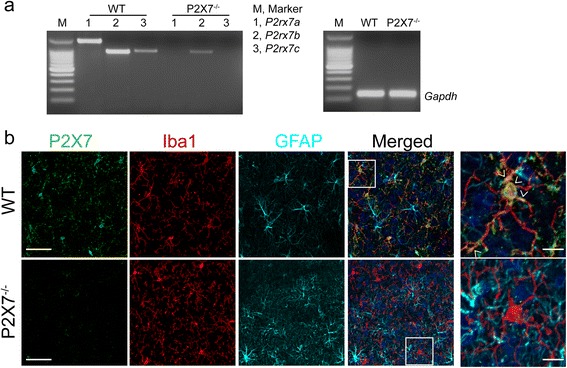
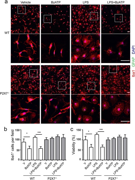
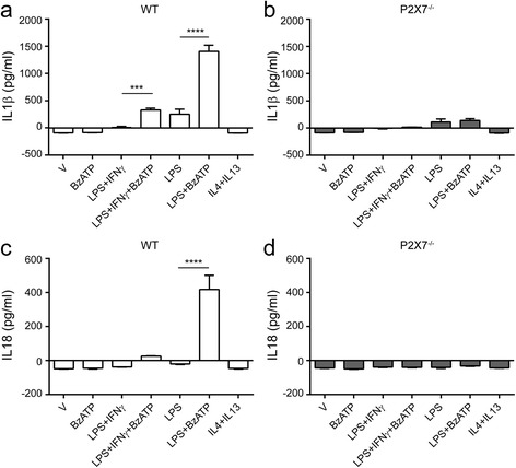
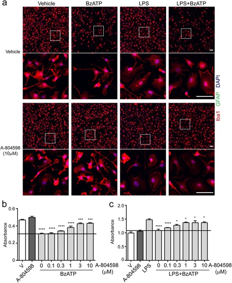
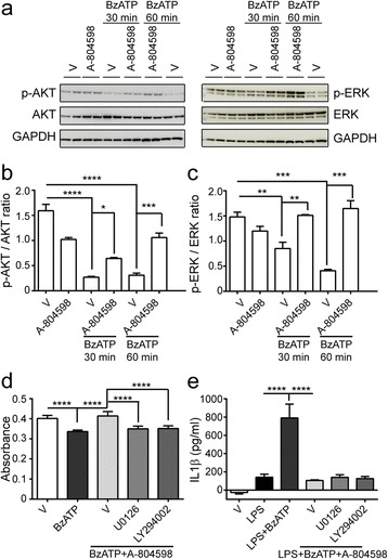
Methods: Primary microglia were isolated from cortices of P0-2 C57BL/6 wild-type or P2X7 knockout (P2X7-/-) mouse pups. Lipopolysaccharide, lipopolysaccharide plus IFNγ, or IL4 plus IL13 were used to polarize microglia to pro-inflammatory or anti-inflammatory states. P2rx7 expression level in resting or activated mouse and human microglia was measured by RNA-sequencing and quantitative real-time PCR. Microglial cell death was measured by cell counting kit-8 and immunocytochemistry, and microglial secretion in wild-type or P2X7-/- microglia was examined by Luminex multiplex assay or ELISA using P2X7 agonist BzATP or P2X7 antagonist A-804598. P2X7 signaling was analyzed by Western blot.

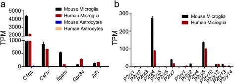
Results: First, we confirmed that P2rx7 is constitutively expressed in mouse and human primary microglia. Moreover, P2rx7 mRNA level was downregulated in mouse microglia under both pro- and anti-inflammatory conditions. Second, P2X7 agonist BzATP caused cell death of mouse microglia, while this effect was suppressed either by P2X7 knockout or by A-804598 under both basal and pro-inflammatory conditions, which suggests the mediating role of P2X7 in BzATP-induced microglial cell death. Third, BzATP-induced release of IL1 family cytokines including IL1α, IL1β, and IL18 was blocked in P2X7-/- microglia or by A-804598 in pro-inflammatory microglia, while the release of other cytokines/chemokines was independent of P2X7 activation. These findings support the specific role of P2X7 in IL1 family cytokine release. Finally, P2X7 activation was discovered to be linked to AKT and ERK pathways, which may be the underlying mechanism of P2X7 functions in microglia.

Conclusions: These results reveal that P2X7 mediates BzATP-induced microglial cell death and specific release of IL1 family cytokines, indicating the important role of P2X7 in neuroinflammation and implying the potential of targeting P2X7 for the treatment of neuroinflammatory disorders.

Keywords: Cell death; Cytokine; Microglia; Neuroinflammation; P2X7.


Figures

Fig. 1
Fig. 1
P2RX and P2RY family expression in microglia. a mRNA levels of microglial marker genes were measured by RNA-seq in human and mouse microglia and astrocytes. b mRNA levels of seven P2rx subtypes and eight P2ry subtypes were measured by RNA-seq in both human and mouse microglia. TPM, transcripts per kilobase million. Data are shown as mean + SD, n = 4
Fig. 2
Fig. 2
Regulation of P2X7 expression in microglia. a mRNA levels of Nos2, Arg1, and P2rx7 in mouse microglia under indicated conditions (100 ng/ml LPS plus 10 ng/ml IFNγ, 10 ng/ml IL4 plus 10 ng/ml IL13, or 100 ng/ml LPS) for 24 h were evaluated by RNA-seq. Data are shown as mean + SD, n = 4. b P2rx7 mRNA levels in mouse microglia exposed to 100 ng/ml LPS plus 10 ng/ml IFNγ, 10 ng/ml IL4 plus 10 ng/ml IL13, 100 ng/ml LPS, or 380 μM BzATP at indicated time durations were measured by quantitative real-time PCR. P2rx7 mRNA level was normalized to Gapdh and presented as fold change compared to vehicle. All experiments were carried out in triplicate and repeated twice independently. Statistical analysis was performed by comparing each condition with vehicle. One-way ANOVA followed by Dunnett’s post hoc test. *P < 0.05; **P < 0.01; ***P < 0.001; ****P < 0.0001
Fig. 3
Fig. 3
P2X7 expression in P2X7−/− mouse brain. a Representative images of gel analysis of reverse transcription PCR products for P2rx7 spliced variants from WT and P2X7−/− microglia. DNA ladder is at 100 bp intervals. Gapdh was used as an internal loading control. b Representative images of immunofluorescence for P2X7 (green), Iba1 (red), and GFAP (cyan) in the cortex of WT and P2X7−/− mice. Nuclei were counterstained with Hoechst (blue). Selected white boxes were zoomed in right panels. Arrowhead indicates representative colocalization of P2X7 and Iba1. Scale bar, 50 μm and 10 μm (zoomed images)
Fig. 4
Fig. 4
P2X7 is necessary for BzATP-induced microglial cell death. Microglia from WT and P2X7−/− mice were primed with or without 100 ng/ml LPS for 22 h following treatment of 380 μM BzATP for additional 4.5 h. a Representative images of double immunofluorescence for GFAP (green) and Iba1 (red) under indicated conditions. Nuclei were counterstained with DAPI (blue). White box indicates position of high magnification images shown below. Scale bar, 50 μm. b Iba1+ microglia quantified by counting at least 3 randomly selected fields under each condition. c Cell viability was calculated by normalizing to vehicle-treated control. Data are shown as mean + SD, n = 3. Experiment was repeated twice independently. One-way ANOVA followed by Tukey’s post hoc test. *P < 0.05; ***P < 0.001
Fig. 5
Fig. 5
Cytokine secretion by WT and P2X7−/− microglia under pro- and anti-inflammatory conditions. Microglia from WT and P2X7−/− mice were treated with 100 ng/ml LPS, 100 ng/ml LPS plus 10 ng/ml IFNγ, or 10 ng/ml IL4 plus 10 ng/ml IL13 for 22 h in the presence or absence of 380 μM BzATP for additional 2 h. ad The secretion of IL1β from WT microglia (a) and P2X7−/− microglia (b), as well as IL18 release from WT microglia (c) and P2X7−/− microglia (d) was assessed by ELISA. Data are shown as mean + SD of triplicates. Experiment repeated twice independently. One-way ANOVA followed by Tukey’s post hoc test. ***P < 0.001; ****P < 0.0001
Fig. 6
Fig. 6
P2X7 antagonist attenuates BzATP-induced microglial cell death. Microglia were pre-treated with different concentrations of P2X7 antagonist A-804598 for 1 h before exposure to 100 ng/ml LPS for 22 h in the absence or presence of 380 μM BzATP for additional 4.5 h. a Immunocytochemical images of microglia with or without P2X7 antagonist treatment under conditions of BzATP alone, LPS alone, or LPS plus BzATP. White box indicates position of high magnification images shown below. Iba1 as microglial marker shown in red, GFAP as astrocyte marker shown in green, DAPI stains nuclei. Scale bar, 50 μm. b The number of microglia treated by BzATP after pre-treatment with different concentrations of A-804598 quantified by CCK-8 kit. c The number of LPS-primed microglia after pre-treatment with different concentrations of A-804598 in the presence of BzATP quantified by CCK-8 kit. Data are shown as mean + SD. Experiment was done in six replicates and repeated three times independently. Statistical analysis was performed between vehicle- and BzATP-treated groups in b, between LPS- and BzATP-treated groups in c. One-way ANOVA followed by Tukey’s post hoc test. *P < 0.05; ***P < 0.001; ****P < 0.0001
Fig. 7
Fig. 7
Secretome of microglia treated with P2X7 antagonist upon pro-inflammatory stimulation. Microglia were incubated with 1 μM P2X7 antagonist A-804598 for an hour before exposure to 100 ng/ml LPS or 100 ng/ml LPS plus 10 ng/ml IFNγ for 22 h in the presence or absence of 380 μM BzATP for additional 2 h. a Heat map and hierarchical clustering of detectable 30 proteins released from microglia measured by Luminex multiplex. be Histogram of IL1β (b), IL1α (c), TNFα (d), and IL6 (e) release obtained from a. Data are shown as mean + SD of triplicates. Experiment repeated twice independently. One-way ANOVA followed by Tukey’s post hoc test. ****P < 0.0001
Fig. 8
Fig. 8
AKT and ERK pathways are involved in BzATP-induced P2X7 activation. ac BzATP was applied to microglial culture for 30 min or 1 h in the absence or presence of A-804598. a Western blots were probed with antibodies against p-AKT and p-ERK. b, c Quantification of p-AKT (b) and p-ERK (c) signal intensity normalized to AKT and ERK, respectively. d Microglial cell death was assessed with the treatments of 50 μM U0126 or 1 μM LY294002 in the presence of 10 μM A-804598 for 1 h prior to 380 μM BzATP stimulation for additional 4.5 h. e Microglia were primed by 100 ng/ml LPS for 22 h followed by 50 μM U0126 or 1 μM LY294002 treatment in the presence of 10 μM A-804598 1 h before 380 μM BzATP stimulation for additional 2 h. IL1β release was then conducted by ELISA. Experiment was repeated at least twice independently. One-way ANOVA followed by Tukey’s post hoc test. *P < 0.05; **P < 0.01; ***P < 0.001; ****P < 0.0001

References

    1. Ransohoff RM, El Khoury J. Microglia in health and disease. Cold Spring Harb Perspect Biol. 2015;8(1):a020560. doi: 10.1101/cshperspect.a020560. - DOI - PMC - PubMed
    1. Bhattacharya A, Derecki NC, Lovenberg TW, Drevets WC. Role of neuro-immunological factors in the pathophysiology of mood disorders. Psychopharmacology. 2016;233(9):1623–1636. doi: 10.1007/s00213-016-4214-0. - DOI - PubMed
    1. Ransohoff RM, Perry VH. Microglial physiology: unique stimuli, specialized responses. Annu Rev Immunol. 2009;27:119–145. doi: 10.1146/annurev.immunol.021908.132528. - DOI - PubMed
    1. Schafer DP, Stevens B. Microglia function in central nervous system development and plasticity. Cold Spring Harb Perspect Biol. 2015;7(10):a020545. doi: 10.1101/cshperspect.a020545. - DOI - PMC - PubMed
    1. Nimmerjahn A, Kirchhoff F, Helmchen F. Resting microglial cells are highly dynamic surveillants of brain parenchyma in vivo. Science. 2005;308(5726):1314–1318. doi: 10.1126/science.1110647. - DOI - PubMed

Substances

LinkOut - more resources