Review
. 2017 Sep;16(9):730-740.
doi: 10.1016/S1474-4422(17)30196-5. Epub 2017 Jul 14.

Detection, risk factors, and functional consequences of cerebral microinfarcts

Affiliations
Review

Detection, risk factors, and functional consequences of cerebral microinfarcts

Susanne J van Veluw et al. Lancet Neurol. 2017 Sep.

Abstract

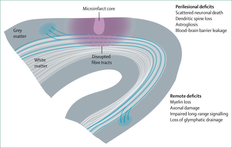
Cerebral microinfarcts are small lesions that are presumed to be ischaemic. Despite the small size of these lesions, affected individuals can have hundreds to thousands of cerebral microinfarcts, which cause measurable disruption to structural brain connections, and are associated with dementia that is independent of Alzheimer's disease pathology or larger infarcts (ie, lacunar infarcts, and large cortical and non-lacunar subcortical infarcts). Substantial progress has been made with regard to understanding risk factors and functional consequences of cerebral microinfarcts, partly driven by new in-vivo detection methods and the development of animal models that closely mimic multiple aspects of cerebral microinfarcts in human beings. Evidence from these advances suggests that cerebral microinfarcts can be manifestations of both small vessel and large vessel disease, that cerebral microinfarcts are independently associated with cognitive impairment, and that these lesions are likely to cause damage to brain structure and function that extends beyond their actual lesion boundaries. Criteria for the identification of cerebral microinfarcts with in-vivo MRI are provided to support further studies of the association between these lesions and cerebrovascular disease and dementia.


Figures

Figure 1
Figure 1. Haematoxylin and eosin-stained sections of cerebral microinfarcts on neuropathological examination
(A) An acute microinfarct (arrows) in the midfrontal cortex and a red hypoxic neuron (inset; arrow). (B) A chronic slit-like microinfarct with puckering in the inferior parietal cortex (arrows). (C) A chronic microinfarct with cavitation (arrow) in the inferior parietal cortex. (D) A chronic microinfarct with haemosiderin deposits in the inferior parietal cortex (inset; arrows).
Figure 2
Figure 2. Incidental small diffusion-weighted imaging lesion
(A) DWI 1·5T MRI shows a hyperintense subcortical lesion (arrow). Follow-up imaging at 3 months shows that the lesion (arrows) is still visible, appearing hypointense on T1 (B) and hyperintense on FLAIR (C). DWI=diffusion-weighted imaging. FLAIR=fluid-attenuated inversion recovery. T1=T1-weighted MRI.
Figure 3
Figure 3. Durable cortical cerebral microinfarcts and mimics on structural MRI
(A) Schematic representation of a cortical cerebral microinfarct (left schematic lesion), which is hypointense on T1, hyperintense on T2 and FLAIR, and isointense on T2*-weighted images. The right schematic lesion (arrow) represents a cortical cerebral microinfarct with cavitation, which appears hypointense on FLAIR with a hyperintense rim. (B) Examples of a cortical cerebral microinfarct (arrows) without cavitation imaged in the same individual on the same day using 7T and 3T MRI. (C) Examples of common cerebral microinfarct mimics, such as cerebral microbleeds (arrows), which are distinct from cerebral microinfarcts because they appear hypointense on T2*-weighted MRI (inset); enlarged perivascular spaces (arrow), which are distinct from cortical cerebral microinfarcts because they are located in juxtacortical areas; and blood vessels (arrow), which are distinct from cortical cerebral microinfarcts because they appear hypointense on T2*-weighted MRI and can be traced over several slices (inset; arrows). FLAIR=fluid-attenuated inversion recovery. T1=T1-weighted MRI. T2=T2-weighted MRI. T2*=T2*-weighted MRI.
Figure 4
Figure 4. Modelling microinfarcts in rodent cortex by targeted photothrombotic occlusion of individual penetrating arterioles
(A) Under the guidance of multiphoton imaging, a single penetrating arteriole is visualised with a scanning laser and occluded with a second fixed green laser after intravenous injection of rose bengal (a photosensitising agent). (B) In-vivo multiphoton images of a single penetrating arteriole, before and after occlusion. (C) A cortical microinfarct visualised using in-vivo T2-weighted MRI. Despite a large difference in brain size, rodent microinfarcts are similar in absolute size to human microinfarcts because microvascular topology is relatively well conserved between species.,
Figure 5
Figure 5. Functional deficits in tissues beyond the non-viable microinfarct core
Perilesional deficits are caused by secondary effects of ischaemic injury, such as spreading depression, blood–brain barrier disruption, and neuroinflammation. Remote deficits arise when microinfarcts damage white matter fibres, or occur in areas that are connected to, and are crucial for, the function of other brain regions

References

    1. Smith EE, Schneider JA, Wardlaw JM, Greenberg SM. Cerebral microinfarcts: the invisible lesions. Lancet Neurol. 2012;11:272–82. - PMC - PubMed
    1. Brundel M, De Bresser J, Van Dillen JJ, Kappelle LJ, Biessels GJ. Cerebral microinfarcts: a systematic review of neuropathological studies. J Cereb Blood Flow Metab. 2012;32:425–36. - PMC - PubMed
    1. Westover MB, Bianchi MT, Yang C, Schneider JA, Greenberg SM. Estimating cerebral microinfarct burden from autopsy samples. Neurology. 2013;13:1–5. - PMC - PubMed
    1. Auriel E, Westover MB, Bianchi MT, et al. Estimating total cerebral microinfarct burden from diffusion-weighted imaging. Stroke. 2015;46:2129–35. - PMC - PubMed
    1. Sonnen JA, Larson EB, Crane PK, et al. Pathological correlates of dementia in a longitudinal, population-based sample of aging. Ann Neurol. 2007;62:406–13. - PubMed

Publication types