Review
. 2018 Apr 2;10(4):a033118.
doi: 10.1101/cshperspect.a033118.

Clinical Neurology and Epidemiology of the Major Neurodegenerative Diseases

Affiliations
Review

Clinical Neurology and Epidemiology of the Major Neurodegenerative Diseases

Michael G Erkkinen et al. Cold Spring Harb Perspect Biol. .

Abstract

Neurodegenerative diseases are a common cause of morbidity and cognitive impairment in older adults. Most clinicians who care for the elderly are not trained to diagnose these conditions, perhaps other than typical Alzheimer's disease (AD). Each of these disorders has varied epidemiology, clinical symptomatology, laboratory and neuroimaging features, neuropathology, and management. Thus, it is important that clinicians be able to differentiate and diagnose these conditions accurately. This review summarizes and highlights clinical aspects of several of the most commonly encountered neurodegenerative diseases, including AD, frontotemporal dementia (FTD) and its variants, progressive supranuclear palsy (PSP), corticobasal degeneration (CBD), Parkinson's disease (PD), dementia with Lewy bodies (DLB), multiple system atrophy (MSA), and Huntington's disease (HD). For each condition, we provide a brief overview of the epidemiology, defining clinical symptoms and diagnostic criteria, relevant imaging and laboratory features, genetics, pathology, treatments, and differential diagnosis.


Figures

Figure 1.
Figure 1.
Magnetic resonance imaging (MRI) of classic Alzheimer’s disease (AD). Coronal T1-weighted brain MRI of a 72-year-old right-handed man with memory problems for at least 4 years showing bilateral hippocampal, and less severe frontal and temporal cortical, atrophy. Orientation is radiologic (right side of figure is left side of brain). (From Sha and Rabinovici 2016, reprinted, with permission, from John Wiley and Sons.)
Figure 2.
Figure 2.
Magnetic resonance imaging (MRI) in three variants of frontotemporal dementia (FTD). T1-weighted brain MRIs in behavioral variant FTD (bvFTD) (A), semantic variant primary progressive aphasia (svPPA) (B), and nonfluent variant (nfvPPA) (C). (A) A 55-year-old woman with a 4-year history of bvFTD with a score of 27/30 on the mini-mental status examination (MMSE) showing an axial, coronal, and sagittal (right side) MRI with significant bilateral (right more than left) frontal atrophy. (B) A 61-year-old man with svPPA showing symptoms for 1.5 years that included forgetting the names of friends and the names and knowledge of common objects. He also showed difficulty with planning, multitasking, and marked rigidity of daily routines. MRI shows severe left temporal pole atrophy. (C) A 74-year-old man with 2 years of progressive word-finding difficulty, slowed and effortful speech, phonemic paraphasias, and speech apraxia. MRI shows left insular and perisylvian atrophy consistent with nfvPPA. Orientation of coronal and axial MRIs are radiologic. (Images courtesy of Dr. David Perry.)
Figure 3.
Figure 3.
“Hot-cross-bun” sign of multiple system atrophy (MSA) of the cerebellar-predominant subtype (MSA-C). Axial T2-weighted magnetic resonance imaging (MRI) of the brain of a 52-year-old patient four years after the onset of MSA-C shows a cruciform T2 hyperintensity in the pons called the “hot-cross-bun sign” (indicated by arrows). Cerebellum shows atrophy. Orientation is radiological. Note that this sign is not specific for MSA, as it occurs in other cerebellar degenerative disorders, such as some of the spinocerebellar ataxias.
Figure 4.
Figure 4.
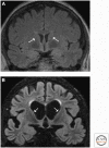
Magnetic resonance imaging (MRI) in healthy normal subject versus patient with Huntington’s disease (HD). Coronal T2-weighted fluid-attenuated inversion recovery (FLAIR) MRI in (A) a healthy control subject and (B) an HD patient. (A) A 68-year-old healthy individual shows normal caudate size (indicated by arrows). (B) A 67-year-old patient with moderate stage HD, 7 years after onset shows diffuse cortical atrophy with disproportionate caudate atrophy (arrows) and corresponding enlargement of the lateral ventricles. Orientation is radiological.

References

    1. Aarsland D, Ballard C, Walker Z, Bostrom F, Alves G, Kossakowski K, Leroi I, Pozo-Rodriguez F, Minthon L, Londos E. 2009. Memantine in patients with Parkinson’s disease dementia or dementia with Lewy bodies: A double-blind, placebo-controlled, multicentre trial. Lancet Neurol 8: 613–618. - PubMed
    1. Ahmed Z, Asi YT, Sailer A, Lees AJ, Houlden H, Revesz T, Holton JL. 2012. The neuropathology, pathophysiology and genetics of multiple system atrophy. Neuropathol Appl Neurobiol 38: 4–24. - PubMed
    1. Alladi S, Xuereb J, Bak T, Nestor P, Knibb J, Patterson K, Hodges JR. 2007. Focal cortical presentations of Alzheimer’s disease. Brain 130: 2636–2645. - PubMed
    1. Alves G, Larsen JP, Emre M, Wentzel-Larsen T, Aarsland D. 2006. Changes in motor subtype and risk for incident dementia in Parkinson’s disease. Mov Disord 21: 1123–1130. - PubMed
    1. Alzheimer’s Association. 2016. 2016 Alzheimer’s disease facts and figures. Alzheimers Dement 12: 459–509. - PubMed

Publication types

MeSH terms