. 2017 Sep 15;77(18):5118-5128.
doi: 10.1158/0008-5472.CAN-16-0342. Epub 2017 Jul 17.

CTLA4 Promotes Tyk2-STAT3-Dependent B-cell Oncogenicity

Affiliations

CTLA4 Promotes Tyk2-STAT3-Dependent B-cell Oncogenicity

Andreas Herrmann et al. Cancer Res. .

Abstract

CTL-associated antigen 4 (CTLA4) is a well-established immune checkpoint for antitumor immune responses. The protumorigenic function of CTLA4 is believed to be limited to T-cell inhibition by countering the activity of the T-cell costimulating receptor CD28. However, as we demonstrate here, there are two additional roles for CTLA4 in cancer, including via CTLA4 overexpression in diverse B-cell lymphomas and in melanoma-associated B cells. CTLA4-CD86 ligation recruited and activated the JAK family member Tyk2, resulting in STAT3 activation and expression of genes critical for cancer immunosuppression and tumor growth and survival. CTLA4 activation resulted in lymphoma cell proliferation and tumor growth, whereas silencing or antibody-blockade of CTLA4 in B-cell lymphoma tumor cells in the absence of T cells inhibits tumor growth. This inhibition was accompanied by reduction of Tyk2/STAT3 activity, tumor cell proliferation, and induction of tumor cell apoptosis. The CTLA4-Tyk2-STAT3 signal pathway was also active in tumor-associated nonmalignant B cells in mouse models of melanoma and lymphoma. Overall, our results show how CTLA4-induced immune suppression occurs primarily via an intrinsic STAT3 pathway and that CTLA4 is critical for B-cell lymphoma proliferation and survival. Cancer Res; 77(18); 5118-28. ©2017 AACR.


Figures

Figure 1
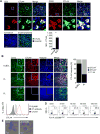
Figure 1. CTLA4 expression and function by B cell lymphoma cells
(A) Immunohistochemical staining followed by confocal microscopy analyses showing CTLA4 expression in CD3+ T cells and CD20+ cells in human B cell lymphoma tissues. Indicated areas (white boxes) magnified. Scale, 50 μm (upper panels). CTLA4 expression in normal human lymph node vs. lymph node with B cell lymphoma, shown by confocal images and quantification (lower panels). (B) Representative microscopic images showing elevated CTLA4 expression by human B cell lymphoma DLBCL and FL (left) tumor sections. Quantified frequency of CTLA4 expression in all of the analyzed patient tumor biopsies (n=11 for both tumor types) (right). Scale, 50 μm. (C) CTLA4 surface expression by human B cell lymphoma cell line Ly3 assessed by flowcytometry. (D) Flowcytometry and (E) confocal microscopy showing cellular internalization of soluble CD86 by Ly3 cells. Scale, 10 μm.
Figure 2
Figure 2. CTLA4 contributes to CD86 cellular internalization
(A) CTLA4-positive A20 B cell lymphoma cells uptake CD86 from APCs. CD86-mCherry expressing RAW macrophages were co-cultured with CFSE+ A20 cells. Cellular internalization of full-length CD86-mCherry by A20 cells was visualized by confocal microscopy. Scale, 10 μm. (B) Flowcytometric quantitative analysis showing CD86-mCherry cellular internalization expressed by RAW macrophages (upper panels) or dendritic cells (lower panels) by CFSE+ A20 cells. (C) Flowcytometric analyses of CD80, CD86, CD28 and CTLA4 in murine A20 B cell lymphoma cells. (D) CTLA4 blockade reduces sCD86 internalization by human B cell lymphoma Ly3 (upper panels) and CTLA4+ Raji cells (lower panels) assessed by flowcytometry.
Figure 3
Figure 3. Tyrosine 218 in CTLA4-mediates ligand internalization in B cells
Mouse pre-B cells stably expressing hCTLA4-GFP constructs, with indicated tyrosine mutations, were used to assess internalization of fluorescently labeled human sCD86. Top, schematic structure of hCTLA4 with or without mutations at tyrosine phophorylation sites. Red line indicates mutations site. Bottom, representative flowcytometry analyses showing internalization of sCD86 by wild-type and mutated hCTLA4. The experiments were repeated three times with similar results.
Figure 4
Figure 4. CD86-CTLA4 intracellular signaling activates Tyk2 and STAT3 in B cell lymphoma cells
(A) CD86-CTLA4 engagement immediately triggers CTLA4 tyrosine phosphorylation and recruitment of STAT3 in Ly3 cells. Ly3 tumor cells were treated with sCD86 followed by immunoprecipitation with CTLA4 antibody and Western blotting to detect pTyr-CTLA4 and STAT3. (B) Tyk2, but not JAK1, 2 or 3, undergoes tyrosine phosphorylation upon exposure to sCD86. (C) Exposure of Ly3 cells to sCD86 results in recruitment of Tyk2 by CTLA4 as assessed by co-immunoprecipitation and Western blotting. (D) (E) CD86 induces immediate STAT3 tyrosine phosphorylation as shown by flowcytometry (D), and by EMSA employing a radiolabeled dsDNA oligo (SIE) harboring a STAT1 and STAT3 binding consensus sequence (E). *) indicates STAT3 supershift with a STAT3 specific antibody. (F) RT-PCR shows effects of CTLA4 –CD86 engagement on mRNA expression of STAT3 target oncogenic genes (left panels) and immune regulatory genes (right panels) in human B cell lymphoma Ly3 cells, which were stimulated by sCD86 stimulation for 24 h. (G) CTLA4 blockade reduces sCD86 induced STAT3 activation as shown by Western blotting and (H) subsequent effects on STAT3 downstream gene expression assessed by RT-PCR for mRNA in three B cell lymphoma cell lines as indicated SD shown. T-test: *) P < 0.05, **) P < 0.01; ***) P < 0.001.
Figure 5
Figure 5. CD86-CTLA4 engagement promotes B cell lymphoma proliferation and growth via Tyk2-STAT3
(A) CD86 on APCs stimulates lymphoma cell proliferation. CD86-mCherry expressing RAW macrophages (left panels) or DC2.4 cells (right panels) were incubated with CFSE+ A20 lymphoma cells followed by flowcytometry to assess dividing A20 cells (upper panels). Highly proliferative CFSE-low versus non-proliferative CFSE-high A20 cells were compared for CD86-mCherry internalization (lower panels). (B) CTLA4 antibody-blockade significantly reduced A20 lymphoma growth in syngeneic mice. (C) CTLA4 blockade in vivo significantly decreased Ki67+ proliferative activity. Scale for confocal microscopy, 100 μm. Ki67 mean fluorescence quantified. (D) CTLA4 knockdown in Ly3 B cell lymphoma reduced tumor growth in vivo in a xenograft model and decreased Ki67 expression in tumor tissue analyzed by confocal microscopy (E). Scale, 50 μm. (F) Blocking CTLA4 significantly delayed human B cell lymphoma growth in immunodeficient mice. (G) Blocking CTLA4 in vivo reduced Tyk2 activation and STAT3 recruitment in human lymphoma, as shown by Western blotting using tumor homogenates from the tumors shown in (F). (H) CTLA4 blockade in human B cell lymphoma in vivo inhibits lymphoma oncogenesis, indicated by changes in levels of CD31, Ki67, and cleaved caspase 3+ in the lymphoma tumors. Confocal microscopy scale, 100 μm and 50 μm. CD31, Ki67, and cleaved caspase 3 mean fluorescence quantified. SD shown. T-test: *) P < 0.05 **) P < 0.01; ***) P < 0.001.
Figure 6
Figure 6. CTLA4-Tyk2-STAT3 oncogenic signaling is active in tumor-associated B cells
(A) CTLA4-blockade inhibits tumor growth of B16 melanoma in syngeneic mice. SD shown. T-test: *) P < 0.05, **) P < 0.01. (B) Flowcytometric analyses show that CTLA4 antibody blockade inhibits Tyk2 and Stat3 activity as well as expression of c-Myc oncogene in CD19+ B cells isolated from the TDLNs. (C) Reduced c-Myc expression by melanoma infiltrating CD19+ B cells upon CTLA4 blockade was confirmed by confocal microscopy. Scale, 20 μm. (D) In vivo blockade of CTLA4 induces CD8 T cells melanoma infiltration. (E) The tumor- infiltrating CD8 T cells are mostly CD69+. (F) Flowcytometric analyses indicate the effects of CTLA4 blockade on non-malignant B cells from lymph nodes of A20 subcutaneous tumor bearing mice (n=4/cohort).

References

    1. Peggs KS, Quezada SA, Korman AJ, Allison JP. Principles and use of anti-CTLA4 antibody in human cancer immunotherapy. Curr Opin Immunol. 2006;18:206–13. - PubMed
    1. Pardoll DM. The blockade of immune checkpoints in cancer immunotherapy. Nat Rev Cancer. 2012;12:252–64. - PMC - PubMed
    1. Thompson CB, Allison JP. The emerging role of CTLA-4 as an immune attenuator. Immunity. 1997;7:445–50. - PubMed
    1. Crespo J, Sun H, Welling TH, Tian Z, Zou W. T cell anergy, exhaustion, senescence, and stemness in the tumor microenvironment. Curr Opin Immunol. 2013;25:214–21. - PMC - PubMed
    1. Mantovani A. B cells and macrophages in cancer: yin and yang. Nat Med. 2011;17:285–6. - PubMed

Publication types

MeSH terms