. 2017 Aug 1;114(31):8360-8365.
doi: 10.1073/pnas.1707662114. Epub 2017 Jul 17.

Sialylation on O-glycans protects platelets from clearance by liver Kupffer cells

Affiliations

Sialylation on O-glycans protects platelets from clearance by liver Kupffer cells

Yun Li et al. Proc Natl Acad Sci U S A. .

Abstract

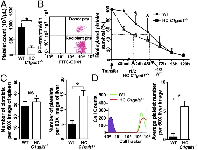
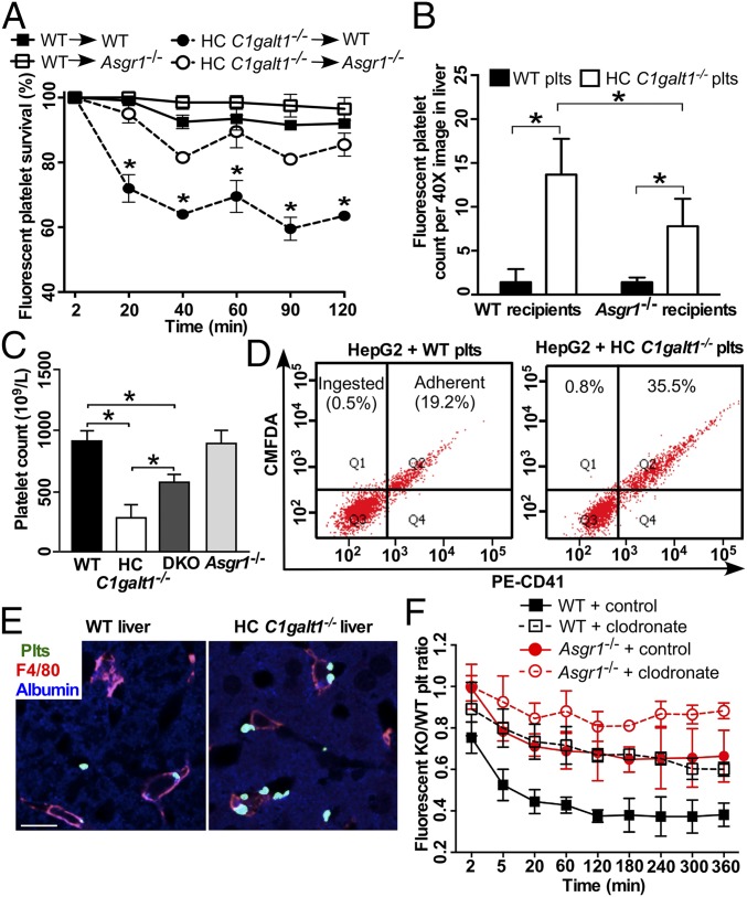
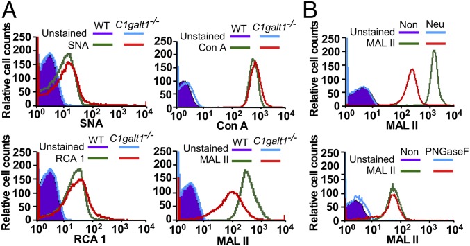
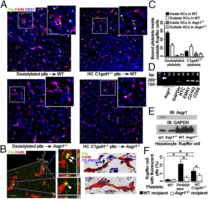
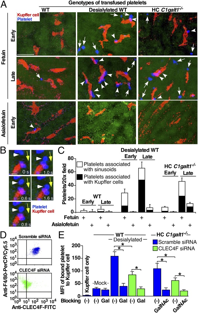
Most platelet membrane proteins are modified by mucin-type core 1-derived glycans (O-glycans). However, the biological importance of O-glycans in platelet clearance is unclear. Here, we generated mice with a hematopoietic cell-specific loss of O-glycans (HC C1galt1-/- ). These mice lack O-glycans on platelets and exhibit reduced peripheral platelet numbers. Platelets from HC C1galt1-/- mice show reduced levels of α-2,3-linked sialic acids and increased accumulation in the liver relative to wild-type platelets. The preferential accumulation of HC C1galt1-/- platelets in the liver was reduced in mice lacking the hepatic asialoglycoprotein receptor [Ashwell-Morell receptor (AMR)]. However, we found that Kupffer cells are the primary cells phagocytosing HC C1galt1-/- platelets in the liver. Our results demonstrate that hepatic AMR promotes preferential adherence to and phagocytosis of desialylated and/or HC C1galt1-/- platelets by the Kupffer cell through its C-type lectin receptor CLEC4F. These findings provide insights into an essential role for core 1 O-glycosylation of platelets in their clearance in the liver.

Keywords: Kupffer cell; O-glycan; clearance; platelet.


Conflict of interest statement

The authors declare no conflict of interest.

Figures

Fig. 1.
Fig. 1.
HC C1galt1−/− mice develop thrombocytopenia with reduced platelet life-span, and HC C1galt1−/− platelets display a greater clearance than WT platelets in the liver. (A) Peripheral platelet count in WT and HC C1galt1−/− mice. Data represent means ± SD. n = 5 mice. *P < 0.05. (B) Representative dot plot shows that 108 biotinylated platelets from WT mice or HC C1galt1−/− mice were adoptively transfused into WT recipients. Peripheral blood samples were taken from recipient mice at 0, 0.3, 24, 48, 72, 96, and 120 h after transfusion. Data represent means ± SD at each time point. n = 4 experiments. *P < 0.05. (C) Bar graphs are quantification of platelets or platelet clusters per 60× image. Data represent mean ± SD. n = 10 images/genotype. *P < 0.05. (D) Flow-cytometric analysis of labeling efficiency of WT and HC C1galt1−/− platelets by CellTracker CMRA (red) and CMFDA (green), respectively. Bar graphs are quantification of number of platelets per 60× field in WT recipients 90 min after the competitive transfusion of labeled WT and HC C1galt1−/− platelets. n = 3 experiments. *P < 0.05.
Fig. 2.
Fig. 2.
The AMR participates in HC C1galt1−/− platelet clearance. (A) Survival percentage of competitively transfused CMRA-labeled WT platelets or CMFDA-labeled HC C1galt1−/− platelets in peripheral blood of either WT or Asgr1−/− recipients. n = 3 experiments. *P < 0.05. (B) Quantification of labeled WT or HC C1galt1−/− platelets in recipient liver sections. Data are mean ± SD. n equals at least five randomly selected 40× microscopic fields. *P < 0.05. (C) Peripheral platelet count of WT and HC C1galt1−/−;Asgr1−/− mice. n = 3 mice/genotype. *P < 0.05. (D) Flow-cytometric analysis of HepG2 cell ingestion of CMFDA-labeled platelets. Platelet adherent to HepG2 cells were identified as CD41 and CMFDA double positive. CMFDA single-positive HepG2 cells suggest ingestion of platelets. The dot plots are representative of five experiments. (E) Representative images of immunostaining of platelets (anti-thrombocyte serum, green), Kupffer cells (F4/80, red), and hepatocytes (albumin, blue) in liver sections. (Scale bar, 10 μm.) (F) The ratio of competitively transfused fluorescent HC C1galt1−/− platelets to WT platelets in peripheral blood of WT and Asgr1−/− recipients with or without Kupffer cell depletion. Data are mean ± SD at each time point. n = 6 mice per group.
Fig. 3.
Fig. 3.
(A) Flow-cytometric analysis of WT and HC C1galt1−/− platelets after being stained with lectin SNA (for α-2,6-linked sialic acids), Con A (for mannose in N-glycans), RCA 1 (for galactose exposure), and MALII (for α-2,3-linked sialic acids). Data represent three experiments. (B) MALII profile of WT platelets with or without neuraminidase (Neu) or PNGase F treatment. Data represent two experiments.
Fig. 4.
Fig. 4.
Desialylated or HC C1galt1−/− platelets are associated with Kupffer cells. (A) Representative confocal microscopic images showing association of CMFDA-labeled platelets with the Kupffer cell (F4/80 staining) in the liver. Anti-CD31 labels sinusoidal endothelial cells. The upper right inlet in each panel shows the enlarged image indicated by the white square. (Scale bar, 10 μm.) (B) A 3D reconstructed image with orthogonal projections of z stacks shows platelets phagocytosed (arrows) by or bound to (arrowhead) the Kupffer cell (Left), 3D-reconstructed images with nucleus staining by TO-PRO3 (blue). Arrow marks the Kupffer cell nucleus (Right). (Scale bar, 5 μm.) (C) Quantification of transfused platelets adherent to or inside Kupffer cells in different recipient liver. Data are mean ± SD. n = 6–10; 20× confocal microscopic fields. (D) RT-PCR analysis of expression of Asgr1 and of macrophage markers. Lane 1, WT liver; lane 2, Asgr1−/− liver; lanes 3–8, sorted WT Kupffer cells. (E) Western blot analysis of AMR levels in hepatocytes or Kupffer cells. (F) Flow-cytometric analysis of F4/80-positive Kupffer cells from WT and Asgr1−/− recipients for association with transfused CMFDA-labeled WT, desialylated, or HC C1galt1−/− platelets.
Fig. 5.
Fig. 5.
Confocal intravital microscopy of recipient livers transfused with WT, desialylated, or HC C1galt1−/− platelets. (A) Representative confocal intravital images of the WT recipient liver at 20-s (early) or 30-min (late) time points after transfusion of platelets labeled with CellTracker (blue). Recipients were administered with fetuin or asialofetuin (i.p.) before platelet transfusions. The Kupffer cells were labeled by an injection of PE-anti-F4/80 (i.v.) before transfusion (red). Arrows indicate transfused platelets associated with sinusoidal endothelium. Arrowheads show Kupffer cells associated with transfused platelets. (Scale bar, 10 μm.) (B) Representative serial images at indicated time frames (in seconds) of a platelet (arrowhead) first rolling on the sinusoid surface and then interacting with a Kupffer cell. Yellow arrows mark the direction of blood flow. (Scale bar, 2 μm.) (C) Quantification of interactions of platelets with liver sinusoid surface and with Kupffer cells (KCs) at different time points after platelet transfusion. n = 5–10 images/time point. Data represent means ± SD from three experiments. (D) Expression of CLEC4F on F4/80-positive Kupffer cells from WT mice transfected with scramble or CLEC4F-specific siRNA measured by flow cytometry. (E) Flow-cytometric analysis of binding of PHK26-labeled mock-treated or sialidase-treated (desialylated) platelets or HC C1galt1−/− platelets to Kupffer cells transfected with either scramble or CLEC4F-specific siRNA, with or without pretreatments with 100 μg/mL Gal or GalNAc polymers. Binding was shown as mean fluorescence intensity (MFI). n = 3 experiments. *P < 0.05.

References

    1. de Gaetano G. Historical overview of the role of platelets in hemostasis and thrombosis. Haematologica. 2001;86:349–356. - PubMed
    1. Bertozzi CC, et al. Platelets regulate lymphatic vascular development through CLEC-2-SLP-76 signaling. Blood. 2010;116:661–670. - PMC - PubMed
    1. Herzog BH, et al. Podoplanin maintains high endothelial venule integrity by interacting with platelet CLEC-2. Nature. 2013;502:105–109. - PMC - PubMed
    1. Morrell CN, Aggrey AA, Chapman LM, Modjeski KL. Emerging roles for platelets as immune and inflammatory cells. Blood. 2014;123:2759–2767. - PMC - PubMed
    1. Harker LA. The kinetics of platelet production and destruction in man. Clin Haematol. 1977;6:671–693. - PubMed

Publication types