. 2017 Aug 1;114(31):8402-8407.
doi: 10.1073/pnas.1703338114. Epub 2017 Jul 17.

Metabolic profiles of exercise in patients with McArdle disease or mitochondrial myopathy

Affiliations

Metabolic profiles of exercise in patients with McArdle disease or mitochondrial myopathy

Nigel F Delaney et al. Proc Natl Acad Sci U S A. .

Abstract

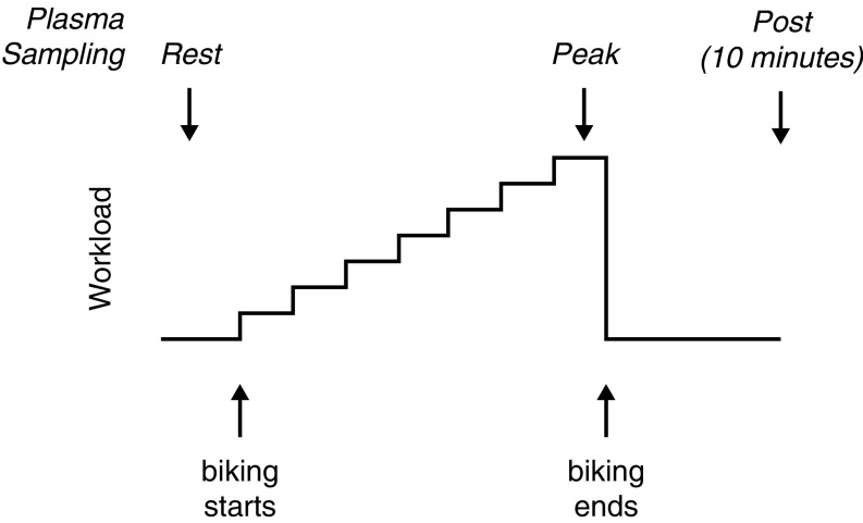
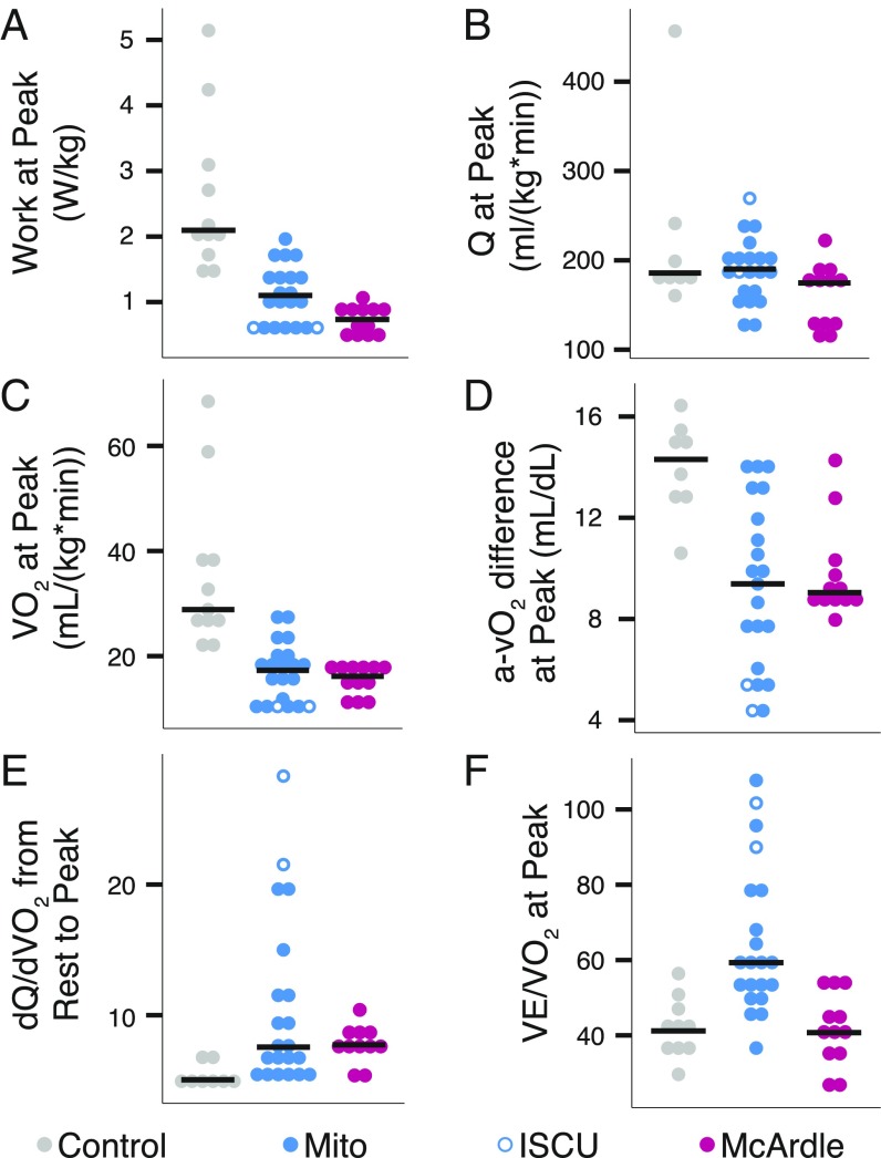
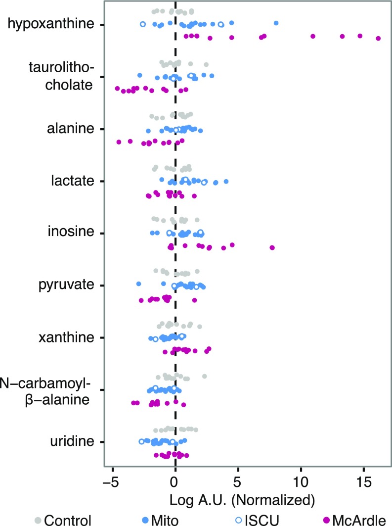
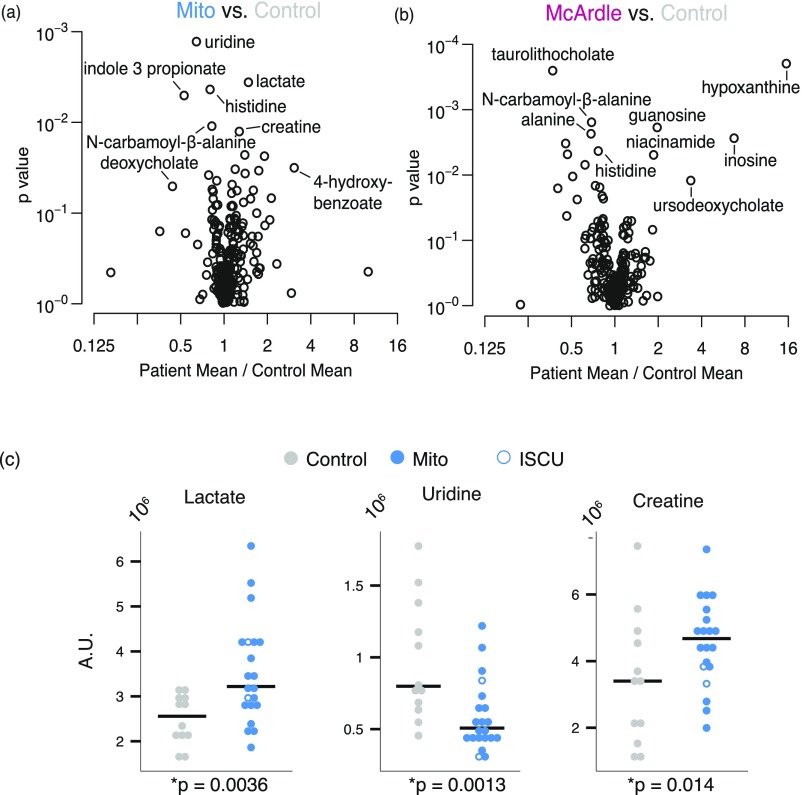
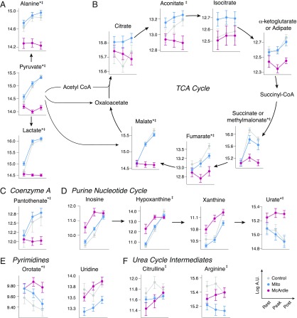
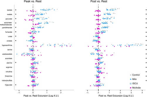
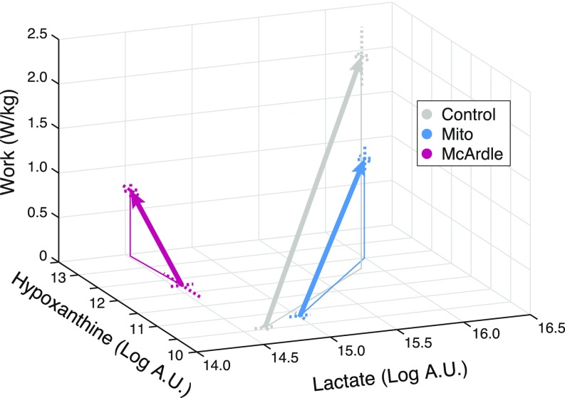
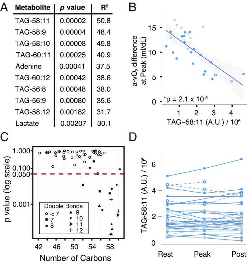
McArdle disease and mitochondrial myopathy impair muscle oxidative phosphorylation (OXPHOS) by distinct mechanisms: the former by restricting oxidative substrate availability caused by blocked glycogen breakdown, the latter because of intrinsic respiratory chain defects. We applied metabolic profiling to systematically interrogate these disorders at rest, when muscle symptoms are typically minimal, and with exercise, when symptoms of premature fatigue and potential muscle injury are unmasked. At rest, patients with mitochondrial disease exhibit elevated lactate and reduced uridine; in McArdle disease purine nucleotide metabolites, including xanthine, hypoxanthine, and inosine are elevated. During exercise, glycolytic intermediates, TCA cycle intermediates, and pantothenate expand dramatically in both mitochondrial disease and control subjects. In contrast, in McArdle disease, these metabolites remain unchanged from rest; but urea cycle intermediates are increased, likely attributable to increased ammonia production as a result of exaggerated purine degradation. Our results establish skeletal muscle glycogen as the source of TCA cycle expansion that normally accompanies exercise and imply that impaired TCA cycle flux is a central mechanism of restricted oxidative capacity in this disorder. Finally, we report that resting levels of long-chain triacylglycerols in mitochondrial myopathy correlate with the severity of OXPHOS dysfunction, as indicated by the level of impaired O2 extraction from arterial blood during peak exercise. Our integrated analysis of exercise and metabolism provides unique insights into the biochemical basis of these muscle oxidative defects, with potential implications for their clinical management.

Keywords: McArdle disease; TCA expansion; exercise physiology; metabolic profiling; mitochondria.


Conflict of interest statement

The authors declare no conflict of interest.

Figures

Fig. S1.
Fig. S1.
Exercise protocol and plasma sampling. Steps show stationary bike workload (measured in Watts) increasing stepwise throughout the exercise test, until subject reached his/her peak workload. Upward arrows denote the beginning and end of exercise; the precise number of minutes exercised varied by individual. Downward arrows denote timing of plasma sample collection.
Fig. S2.
Fig. S2.
Time series of heart rates and workloads. Lines show the heart rate and workload data for each exercise trial in the study. Parameters were recorded at regular intervals shown by dots. Note that to avoid exertional muscle injuries, McArdle patients first acclimated for 15 min at a lower exercise intensity before the start of their trial; this enabled their second wind phenomenon to occur (indicated by the drop in heart rate), which allowed them to achieve workloads more comparable to controls.
Fig. 1.
Fig. 1.
Exercise physiology in patients with mitochondrial myopathy or McArdle disease. Dot plots for individual exercise tests showing (A) work, (B) cardiac output (Q), (C) oxygen uptake (VO2), (D) systemic arteriovenous oxygen difference (a-vO2), (E) the slope of the increase in cardiac output relative to the increase in oxygen utilization during exercise, and (F) ventilation (VE) relative to VO2 at maximal exercise. Horizontal lines denote median value by group. A permutation test showed the patient cohorts were all significantly different from the control group (p < 0.05) except for Mito in B and McArdle group in F.
Fig. 2.
Fig. 2.
Plasma metabolites at rest in patients with mitochondrial myopathy or McArdle disease. Scatterplots of metabolites whose mean levels at rest significantly differed among the three subject groups ordered top to bottom by statistical significance. The measurements are in logged arbitrary units (A.U.) and normalized to the mean and variance of the control group.
Fig. S3.
Fig. S3.
Individual group comparison and validation of biomarkers for mitochondrial disease. (A and B) Volcano plots showing the spectrum of metabolic differences in the pairwise comparisons between (A) Mito patients vs. controls and (B) McArdle patients vs. controls. The x axis shows the ratio of the mean patient value to the mean control value, plotted against the p value for the test of equal means in both groups. (C) Resting measurements of lactate, uridine, and creatine that were identified both in this study and previous studies as distinguishing Mito from control.
Fig. 3.
Fig. 3.
(A–F) Metabolic pathways whose metabolites change with exercise in patients with mitochondrial myopathy or McArdle disease. Metabolite levels during exercise shown for those in pathways found to have significantly different excursions in the post/rest comparison. The measurements are in logged arbitrary units (A.U.). Metabolites with significantly different excursions from rest to peak are indicated by an asterisk (*); those that significantly differ from rest to post are indicated by a double dagger ().
Fig. S4.
Fig. S4.
Exercise-induced changes in plasma metabolites among the three groups. Scatter plots showing metabolites whose average change between exercise time points was not equal across the three groups as described in the main text. Metabolite excursions from rest to peak are shown on the Left, whereas excursions from rest to postexercise are shown on the Right. The 10 metabolites with significantly different changes at peak/rest are starred on the Left, whereas the 17 metabolites with significantly different changes at post/rest are starred on the Right.
Fig. 4.
Fig. 4.
Exercise-induced trajectories for lactate and hypoxanthine as a function of work. Mean values of lactate and hypoxanthine are plotted at rest and at peak. Dashed lines indicate SEs.
Fig. 5.
Fig. 5.
Resting metabolites that are predictive of peak exercise systemic a-vO2 difference in patients with mitochondrial myopathy. (A) Ordered list of the top 10 significant metabolites when the systemic a-vO2 difference at peak was regressed against the metabolite level at rest. (B) Regression result for the most significant metabolite, TAG-58:11. (C) For the 52 TAG species measured, a comparison of how the p value in the regression from A relates to the number of carbons in the TAG species. Symbols show the number of double bonds. (D) TAG-58:11 variation with exercise testing; points connected by a single line represent measurements from the same trial. Symbols and colors as in Fig. 1.
Fig. S5.
Fig. S5.
TAG stability. Each point represents two measurements the measurement of a TAG on separate days for participants who underwent two exercise trials. Points are only shown for the eight TAGs identified in Fig. 5A.

References

    1. Chen Y-T, Kishnani PS, Koeberl D. 2014. Glycogen storage diseases. OMMBID: The Online Metabolic and Molecular Bases of Inherited Diseases, eds Valle D, et al. (McGraw Hill, New York), Chap 71.
    1. McArdle B. Myopathy due to a defect in muscle glycogen breakdown. Clin Sci. 1951;10:13–35. - PubMed
    1. Rumpf KW, et al. Increased ammonia production during forearm ischemic work test in McArdle’s disease. Klin Wochenschr. 1981;59:1319–1320. - PubMed
    1. Haller RG, Vissing J. Spontaneous “second wind” and glucose-induced second “second wind” in McArdle disease: Oxidative mechanisms. Arch Neurol. 2002;59:1395–1402. - PubMed
    1. Taivassalo T, et al. The spectrum of exercise tolerance in mitochondrial myopathies: A study of 40 patients. Brain. 2003;126:413–423. - PubMed

Publication types

MeSH terms