. 2017 Aug 1;114(31):8342-8347.
doi: 10.1073/pnas.1704259114. Epub 2017 Jul 17.

Severe viral respiratory infections in children with IFIH1 loss-of-function mutations

Affiliations

Severe viral respiratory infections in children with IFIH1 loss-of-function mutations

Samira Asgari et al. Proc Natl Acad Sci U S A. .

Abstract

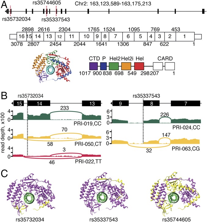
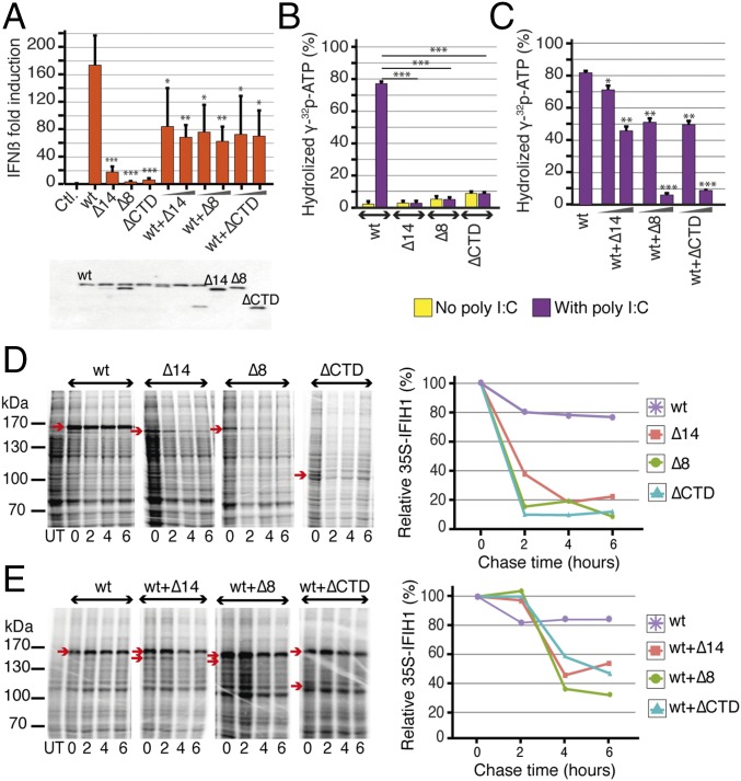
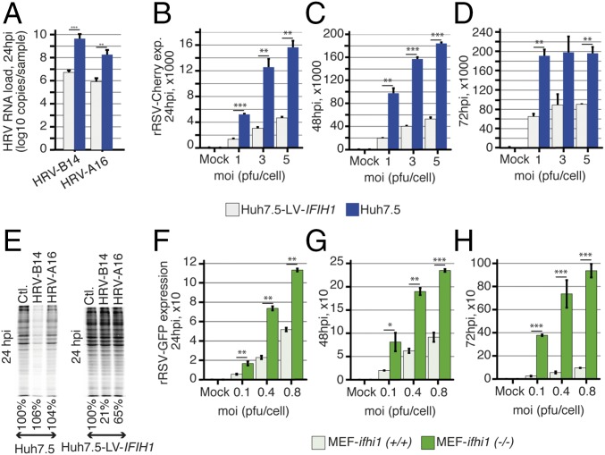
Viral respiratory infections are usually mild and self-limiting; still they exceptionally result in life-threatening infections in previously healthy children. To investigate a potential genetic cause, we recruited 120 previously healthy children requiring support in intensive care because of a severe illness caused by a respiratory virus. Using exome and transcriptome sequencing, we identified and characterized three rare loss-of-function variants in IFIH1, which encodes an RIG-I-like receptor involved in the sensing of viral RNA. Functional testing of the variants IFIH1 alleles demonstrated that the resulting proteins are unable to induce IFN-β, are intrinsically less stable than wild-type IFIH1, and lack ATPase activity. In vitro assays showed that IFIH1 effectively restricts replication of human respiratory syncytial virus and rhinoviruses. We conclude that IFIH1 deficiency causes a primary immunodeficiency manifested in extreme susceptibility to common respiratory RNA viruses.

Keywords: IFIH1; RIG-I-like receptor family; respiratory syncytial virus; rhinovirus; severe pediatric infectious disease.


Conflict of interest statement

The authors declare no conflict of interest.

Figures

Fig. 1.
Fig. 1.
LoF variants identified in IFIH1. Related to SI Appendix, Fig. S1. (A) Schematic representation of IFIH1 DNA, mRNA, and protein. The identified variants are indicated with dashed red lines. Exon boundaries are marked with nucleotide coordinates. Protein domain boundaries are marked with amino acid coordinates. (B) Alternative splicing of IFIH1 associated with rs35732034 and rs35337543 genotypes, as seen in RNA sequencing data. The T allele at rs35732034 leads to skipping of exon 14. The G allele at rs35337543 leads to skipping of exon 8. The Sashimi plots illustrate the genotype-dependent abundance of splice junctions. The number of observed reads spanning the respective splice junctions is indicated on the Bezier curves, which connect exons. (C) Schematic 3D representation of IFIH1 (Protein Data Bank ID code: 4GL2, image produced using UCSC Chimera). The parts of the protein that are predicted to be missing due to rs35732034, rs35744605 and rs35337543 variants are indicated in yellow. Hel, helicase domain; P, pincer.
Fig. 2.
Fig. 2.
Functional characterization of the IFIH1 variants. Related to SI Appendix, Fig. S2. (A) Transfection with IFIH1-wt plasmid (20 ng) results in strong IFNβ induction in 293T cells, whereas transfection of plasmids harboring any of the IFIH1 variants cannot induce IFNβ (240 ng, n = 4). Cotransfection experiments demonstrate an interference of the mutant plasmids with IFIH1-wt on IFNβ induction (20-ng wt plasmid, 20- and 120-ng mutant plasmids, n = 4). Expression levels of the IFIH1 isoforms are shown under the plot in the Western blot gel. *P < 0.05, **P < 0.01, ***P < 0.001. (B) RNA-induced ATPase activity of purified IFIH1-wt protein and alternate IFIH1 isoforms; IFIH1-wt can hydrolyze ATP in the presence of polyI:C, whereas IFIH1-Δ14, IFIH1-Δ8, and IFIH1-ΔCTD lack ATPase activity, with (purple) or without (yellow) polyI:C stimulation. (C) ATPase activity of IFIH1-wt is reduced upon coincubation with the alternate isoforms in a dose-dependent manner (300-ng wt protein, 300- or 600-ng of each alternate isoform, 10-ng polyI:C, n = 2). *P < 0.05, **P < 0.01, ***P < 0.001. (D and E) Protein stability followed by pulse chase in 293T cells expressing IFIH1-wt, IFIH1-Δ14, IFIH1-Δ8, or IFIH1-ΔCTD. Each protein is marked on the gel by an arrow, and relative amounts of proteins are shown in the graphs. IFIH1-wt is more stable than the alternate IFIH1 isoforms, and the stability of IFIH1-wt is reduced upon coexpression with any of the alternate isoforms. Molecular mass markers are shown on the left of each gel (kilodaltons) and the bands corresponding to IFIH1-wt, IFIH1-Δ14, IFIH1-Δ8, or IFIH1-ΔCTD are indicated using a red arrow (2-μg plasmid expressing wt protein, 2 μg of each mutant plasmid, n = 1). Data are represented as mean ± SD; polyI:C, polyinosinic:polycytidylic acid.
Fig. 3.
Fig. 3.
IFIH1 restricts HRV and RSV replication. Related to SI Appendix, Figs. S3 and S4. (A) Real-time PCR of HRV-B14 and HRV-A16 RNA in Huh7.5 and Huh7.5 cells transduced with a lentiviral vector expressing IFIH1 (Huh7.5-LV-IFIH1). HRV-B14 and HRV-A16 replicate more efficiently in the absence of IFIH1 at 24 h postinfection (hpi) (n = 5 for HRV-B14 and n = 2 for HRV-B16). *P < 0.05, **P < 0.01, ***P < 0.001. (B–D) FACS analyses of mCherry expressing recombinant RSV (rRSV-Cherry) in Huh7.5 and Huh7.5-LV-IFIH1 transduced cells at 24, 48, and 72 hpi show that RSV replicates more efficiently in the absence of IFIH1 (n = 2). (E) Cellular proteins labeled with 35S at 24 hpi with both HRV-B14, HRV-A16 showing a much stronger shutoff of cellular protein synthesis in Huh7.5 cells than in Huh7.5-LV-IFIH1 transduced cells, due to higher viral replication. (F–H) FACS analyses of GFP expressing recombinant RSV (rRSV-GFP) in ifih1 knockout MEFs [MEF-ifih1(−/−)] and ifih1 knockout MEFs transduced with a lentiviral vector expressing IFIH1 (MEF-LV-IFIH1). RSV replicates more efficiently in the absence of IFIH1 at 24, 48, and 72 hpi (n = 2). *P < 0.05, **P < 0.01, ***P < 0.001. Data are represented as mean ± SD. GFP, green florescent protein; MEF, mouse embryonic fibroblast; moi, multiplicity of infection; pfu, plaque-forming unit.

References

    1. Meissner HC. Viral bronchiolitis in children. N Engl J Med. 2016;374:62–72. - PubMed
    1. Nair H, et al. Severe Acute Lower Respiratory Infections Working Group Global and regional burden of hospital admissions for severe acute lower respiratory infections in young children in 2010: A systematic analysis. Lancet. 2013;381:1380–1390. - PMC - PubMed
    1. Liu L, et al. Global, regional, and national causes of child mortality in 2000-13, with projections to inform post-2015 priorities: An updated systematic analysis. Lancet. 2015;385:430–440. - PubMed
    1. Jain S, et al. CDC EPIC Study Team Community-acquired pneumonia requiring hospitalization among U.S. children. N Engl J Med. 2015;372:835–845. - PMC - PubMed
    1. Debiaggi M, Canducci F, Ceresola ER, Clementi M. The role of infections and coinfections with newly identified and emerging respiratory viruses in children. Virol J. 2012;9:247. - PMC - PubMed

Publication types

MeSH terms