Severe Hypocalcemia in a Patient with Recurrent Chondrosarcoma
- PMID: 28717079
- PMCID: PMC5548676
- DOI: 10.2169/internalmedicine.56.7884
Severe Hypocalcemia in a Patient with Recurrent Chondrosarcoma
Abstract
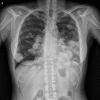
Hypocalcemia is relatively uncommon paraneoplastic syndrome. Only one case of hypocalcemia has been reported in a patient with chondrosarcoma. We herein report a case of a 32-year-old woman with metastatic chondrosarcoma with tetany. Her imaging findings revealed multiple calcific metastatic lesions in the lungs, pancreas, left atrium, and pulmonary vein. A laboratory examination showed hypocalcemia with no evidence of any other disease that could induce hypocalcemia. On the basis of the laboratory and clinical findings, we concluded the etiology of her severe hypocalcemia to be excessive calcium consumption by the tumor itself.
Keywords: chondrosarcoma; hypocalcemia; osteoblastic metastases.
Figures


References
-
- Stewart AF. Hypercalcemia associated with cancer. N Engl J Med 352: 373-379, 2005. - PubMed
-
- Smallridge RC, Wray HL, Schaaf M. Hypocalcemia with osteoblastic metastases in patient with prostate carcinoma. A cause of secondary hyperparathyroidism. Am J Med 71: 184-188, 1981. - PubMed
-
- Tashjian AHJ, Melvin KEW. Medullary carcinoma of the thyroid gland. N Engl J Med 279: 279-283, 1968. - PubMed
-
- Relkin R. Hypocalcemia resulting from calcium accretion by a chondrosarcoma. Cancer 34: 1834-1837, 1974. - PubMed
-
- Fallah-Rad N, Morton AR. Managing hypercalcaemia and hypocalcaemia in cancer patients. Curr Opin Support Palliat Care 7: 265-271, 2013. - PubMed
Publication types
MeSH terms
LinkOut - more resources
Full Text Sources
Other Literature Sources
Medical

