

Cerebrospinal Fluid Rhinorrhea and Subsequent Bacterial Meningitis due to an Atypical Clival Fracture
- PMID: 28717092
- PMCID: PMC5548689
- DOI: 10.2169/internalmedicine.56.8186
Cerebrospinal Fluid Rhinorrhea and Subsequent Bacterial Meningitis due to an Atypical Clival Fracture
Abstract
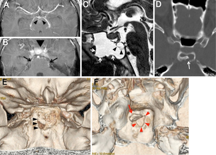
Cases of cerebrospinal fluid (CSF) rhinorrhea due to clival fracture are rare. We present a case of bacterial meningitis with CSF rhinorrhea after a clival fracture. Heavily T2-weighted images showed a bone flap in the thinned clivus and fluid collection in the sphenoid sinus. CSF rhinorrhea developed at 1 month after mild trauma. The fracture may have been caused by the trauma and/or by the pressure gradient between the intracranial CSF space and the sphenoid sinus. A detailed history to identify trauma and an examination to detect bone defects in the skull base are necessary when patients present with bacterial meningitis and persistent rhinorrhea.
Keywords: bacterial meningitis; cerebrospinal fluid; clival fracture; rhinorrhea.
Figures
Comment in
-
Atypical Clival Fracture Due to Minor Trauma and Cerebrospinal Fluid Rhinorrhea.Intern Med. 2017;56(14):1757. doi: 10.2169/internalmedicine.56.8744. Epub 2017 Jul 15. Intern Med. 2017. PMID: 28717069 Free PMC article. No abstract available.
References
-
- Daudia A, Biswas D, Jones NS. Risk of meningitis with cerebrospinal fluid rhinorrhea. Ann Otol Rhinol Laryngol 116: 902-905, 2007. - PubMed
-
- Wang YF, Lirng JF, Fuh JL, Hseu SS, Wang SJ. Heavily T2-weighted MR myelography vs CT myelography in spontaneous intracranial hypotension. Neurology 73: 1892-1898, 2009. - PubMed
-
- Komatsu M, Komatsu F, Cavallo LM, et al. Purely endoscopic repair of traumatic cerebrospinal fluid rhinorrhea from the anterior skull base: case report. Neurol Med Chir (Tokyo) 51: 222-225, 2011. - PubMed
Publication types
MeSH terms
LinkOut - more resources
Full Text Sources
Other Literature Sources
Medical