. 2017 Jul 17;7(1):5588.
doi: 10.1038/s41598-017-05196-1.

3D printed scaffolds of calcium silicate-doped β-TCP synergize with co-cultured endothelial and stromal cells to promote vascularization and bone formation

Affiliations

3D printed scaffolds of calcium silicate-doped β-TCP synergize with co-cultured endothelial and stromal cells to promote vascularization and bone formation

Yuan Deng et al. Sci Rep. .

Abstract

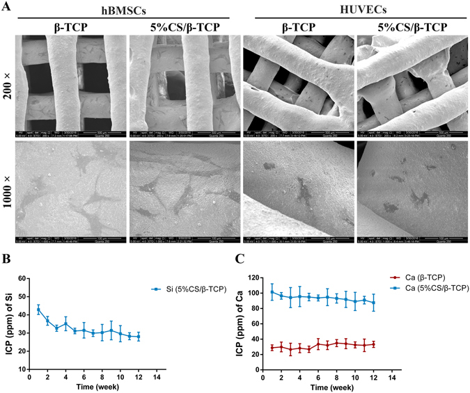
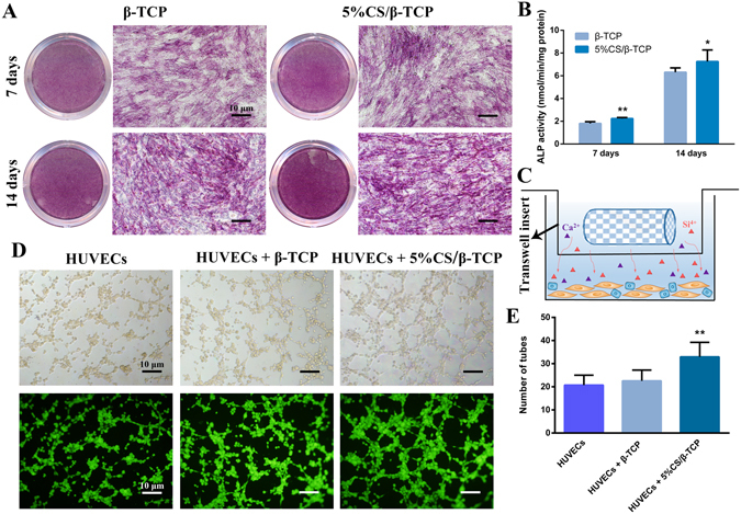
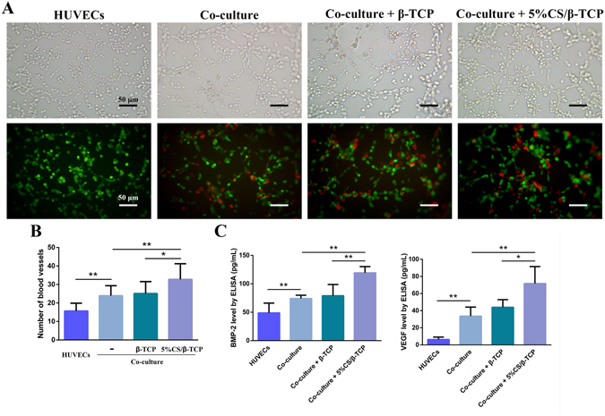
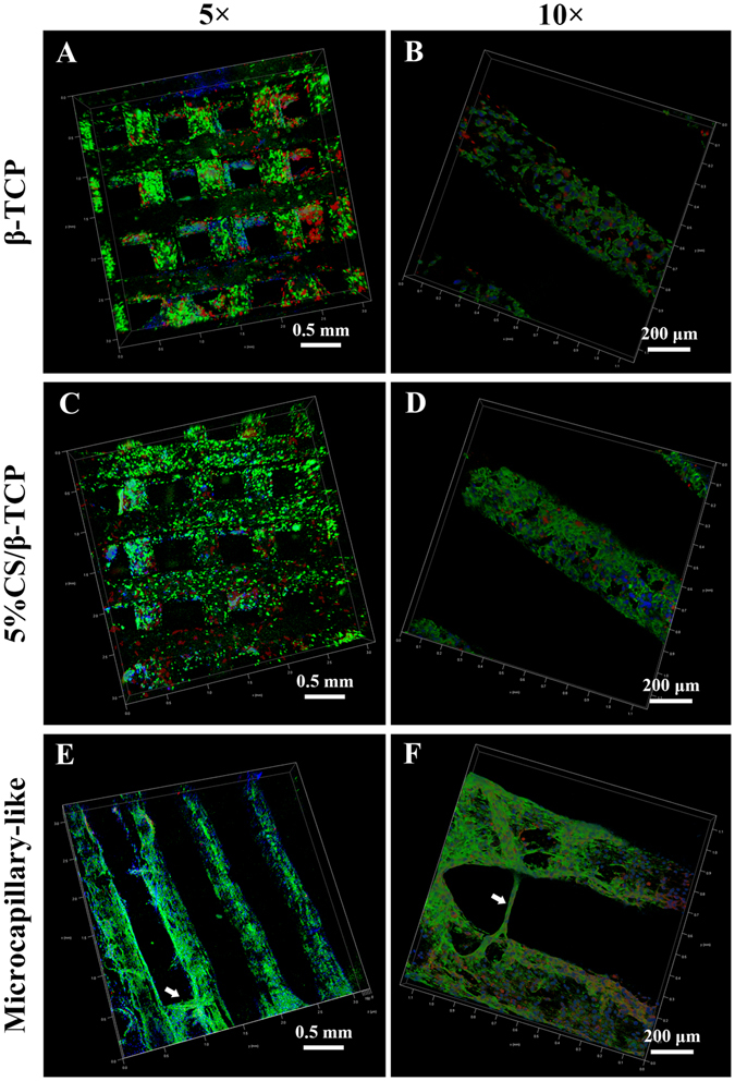
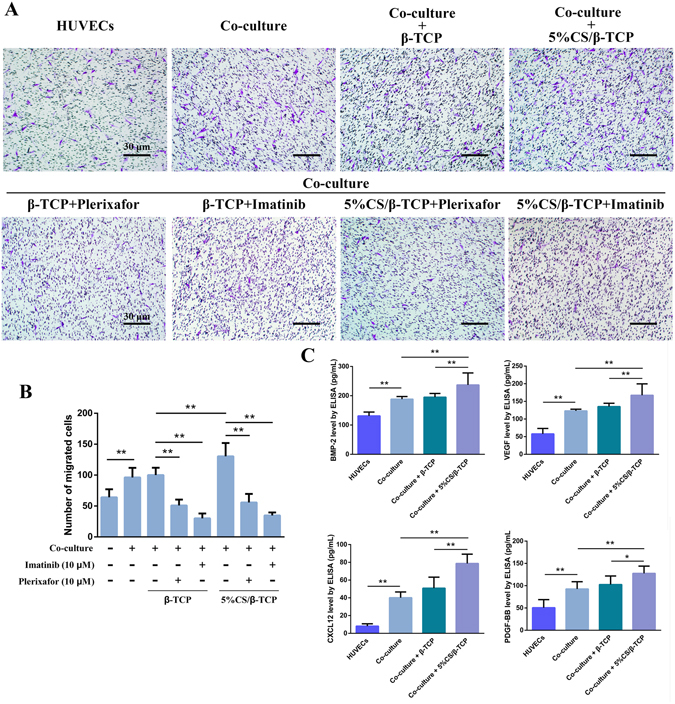
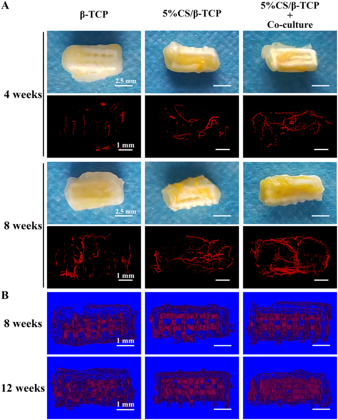
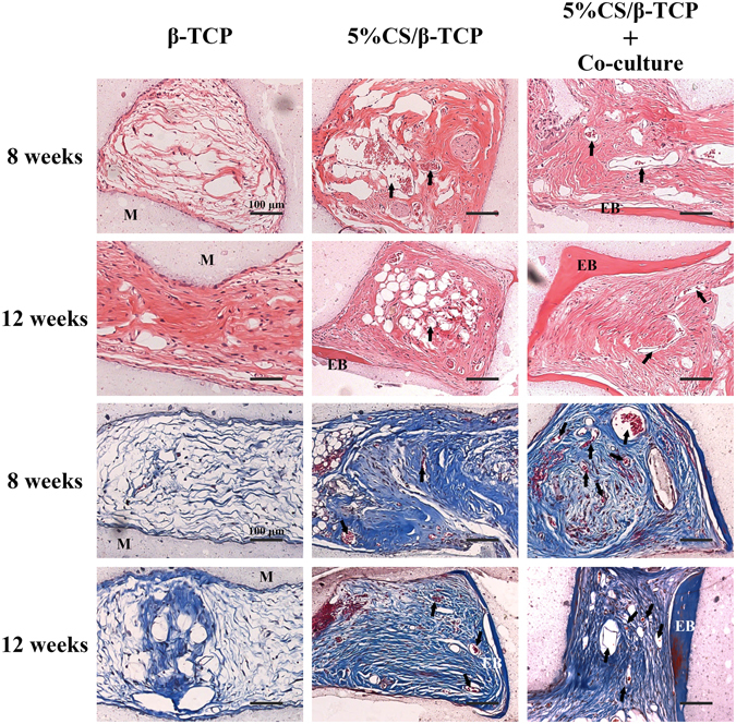
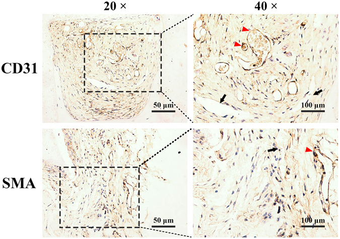
Synthetic bone scaffolds have potential application in repairing large bone defects, however, inefficient vascularization after implantation remains the major issue of graft failure. Herein, porous β-tricalcium phosphate (β-TCP) scaffolds with calcium silicate (CS) were 3D printed, and pre-seeded with co-cultured human umbilical cord vein endothelial cells (HUVECs) and human bone marrow stromal cells (hBMSCs) to construct tissue engineering scaffolds with accelerated vascularization and better bone formation. Results showed that in vitro β-TCP scaffolds doped with 5% CS (5%CS/β-TCP) were biocompatible, and stimulated angiogenesis and osteogenesis. The results also showed that 5%CS/β-TCP scaffolds not only stimulated co-cultured cells angiogenesis on Matrigel, but also stimulated co-cultured cells to form microcapillary-like structures on scaffolds, and promoted migration of BMSCs by stimulating co-cultured cells to secrete PDGF-BB and CXCL12 into the surrounding environment. Moreover, 5%CS/β-TCP scaffolds enhanced vascularization and osteoinduction in comparison with β-TCP, and synergized with co-cultured cells to further increase early vessel formation, which was accompanied by earlier and better ectopic bone formation when implanted subcutaneously in nude mice. Thus, our findings suggest that porous 5%CS/β-TCP scaffolds seeded with co-cultured cells provide new strategy for accelerating tissue engineering scaffolds vascularization and osteogenesis, and show potential as treatment for large bone defects.


Conflict of interest statement

The authors declare that they have no competing interests.

Figures

Figure 1
Figure 1
Cell viability on 5%CS/β-TCP scaffolds. (A) Cell viability, as measured by Cell Counting Kit-8, was comparable between β-TCP scaffolds and other composite scaffolds. Cell viability was significantly inhibited on scaffolds with more than 10% CS. (B) Cell viability of cells around scaffolds was also visualized by Live/Dead Kit, which stains viable cells green and dead cells red. *p < 0.05; **p < 0.01. Scale bar represents 20 μm. M marked materials.
Figure 2
Figure 2
Characterization of 5%CS/β-TCP scaffolds. (A) SEM of HUVECs and hBMSCs seeded on 5%CS/β-TCP scaffolds. Release of (B) Si ions and (C) Ca ions. Scale bar represents 500 μm for 200 X and 100 μm for 1000 X.
Figure 3
Figure 3
5%CS/β-TCP scaffolds stimulate hBMSC osteogenesis and HUVEC angiogenesis in vitro. (A) ALP staining in hBMSCs cultured for 7 and 14 days with β-TCP scaffolds or 5%CS/β-TCP scaffolds in transwell inserts. (B) 5%CS/β-TCP scaffolds stimulated ALP activity at 7 days and 14 days in comparison to pure β-TCP. (C) Schematic representation of transwell experiments. Scaffolds in Transwell inserts (upper chamber), cells in the lower chamber. (D) Tube formation by HUVECs, as observed by light microscopy (top) and Calcein AM staining (bottom), after 4 h on Matrigel with β-TCP or 5%CS/β-TCP scaffolds in transwell inserts. (E) 5%CS/β-TCP scaffolds enhanced tube formation in comparison to β-TCP scaffolds. *p < 0.05; **p < 0.01. Scale bar represents 10 μm.
Figure 4
Figure 4
5%CS/β-TCP scaffolds enhanced angiogenesis in co-cultured HUVECs and hBMSCs. (A) Light microscopy (top) of tube formation on Matrigel, and fluorescence microscopy (bottom) of GFP-labelled HUVECs (green) stimulated for 4 h with β-TCP scaffolds or 5%CS/β-TCP scaffolds in transwell inserts, in the presence or absence of hBMSCs labelled with CM-Dil (red). (B) Co-culture of HUVECs with hBMSCs stimulated tube formation relative to monocultured HUVECs. 5%CS/β-TCP scaffolds further enhanced tube formation in co-cultured cells in comparison to pure β-TCP. (C) BMP-2 and VEGF secretion, as measured by ELISA. *p < 0.05; **p < 0.01. Scale bar represents 50 μm.
Figure 5
Figure 5
Confocal laser scanning microscopy of co-cultured cells on β-TCP and 5%CS/β-TCP scaffolds. Confocal images of HUVECs co-cultured with hBMSCs on (A,B) β-TCP and (C,D) 5%CS/β-TCP scaffolds for 3 days. Viable cells were stained with FITC (green), nuclei were stained with DAPI (blue), and hBMSCs were tracked with CM-Dil (red). (E,F) Confocal images of HUVECs co-cultured with hBMSCs labeled with CM-Dil (red) on 5%CS/β-TCP scaffolds for 10 days. Endothelial cells were stained with CD31 (green), and nuclei were stained with DAPI (blue). White arrows mark CD31-positive microcapillary-like structures.
Figure 6
Figure 6
5%CS/β-TCP scaffolds enhance migration of hBMSCs. (A) Transwell migration assay with β-TCP scaffolds or 5%CS/β-TCP scaffolds, and with 10 μM of the inhibitors plerixafor or imatinib. (B) 5%CS/β-TCP scaffolds enhanced the migration of hBMSCs, an effect blocked by imatinib and plerixafor. (C) BMP-2, VEGF, CXCL12 and PDGF-BB secretion, as measured by ELISA. *p < 0.05; **p < 0.01. Scale bar represents 30 μm.
Figure 7
Figure 7
Analysis of new vessels and new tissue. (A) Macroscopic and micro-CT imaging of new vessels formed at 4 and 8 weeks in β-TCP and 5%CS/β-TCP scaffolds, as well as in 5%CS/β-TCP scaffolds seeded with co-culture cells. (B) Micro-CT analysis of new tissues formed in scaffolds at 8 and 12 weeks. Scale bar represents 2.5 mm for macroscopic and 1 mm for micro-CT.
Figure 8
Figure 8
Histological observation of vessel penetration and ectopic bone formation on β-TCP and 5%CS/β-TCP scaffolds, as well as on 5%CS/β-TCP scaffolds pre-seeded with HUVECs and hBMSCs. Specimens were collected at 8 and 12 weeks, and stained with haematoxylin-eosin and Masson’s trichrome. Arrows indicate new vessels, materials is marked M, and ectopic bone is marked EB. Scale bar represents 100 μm.
Figure 9
Figure 9
Representative immunohistological sections of 5%CS/β-TCP scaffolds pre-seeded with HUVECs and hBMSCs and implanted into nude mice. A few luminal structures contained human CD31 and human SMC (red arrows), but others did not (black arrows) and were solely host-derived.

References

    1. Stegen S, Van GN, Carmeliet G. Bringing new life to damaged bone: The importance of angiogenesis in bone repair and regeneration. Bone. 2015;70:19–27. doi: 10.1016/j.bone.2014.09.017. - DOI - PubMed
    1. Stegen S, van Gastel N, Carmeliet G. Bringing new life to damaged bone: the importance of angiogenesis in bone repair and regeneration. Bone. 2015;70:19–27. doi: 10.1016/j.bone.2014.09.017. - DOI - PubMed
    1. Mankin HJ, Gebhardt Mcjennings LC, Springfield DS, Tomford WW. Long-term results of allograft replacement in the management of bone tumors. Clinical Orthopaedics & Related Research. 1996;324:86–97. doi: 10.1097/00003086-199603000-00011. - DOI - PubMed
    1. Almubarak S, et al. Tissue engineering strategies for promoting vascularized bone regeneration. Bone. 2016;83:197–209. doi: 10.1016/j.bone.2015.11.011. - DOI - PMC - PubMed
    1. Zhai W, et al. Stimulatory effects of the ionic products from Ca-Mg-Si bioceramics on both osteogenesis and angiogenesis in vitro. Acta biomaterialia. 2013;9:8004–8014. doi: 10.1016/j.actbio.2013.04.024. - DOI - PubMed

Publication types

MeSH terms

LinkOut - more resources