. 2016 Aug;20(4):212-224.
doi: 10.1016/j.cossms.2016.03.002. Epub 2016 Mar 28.

Biomaterials for 4D stem cell culture

Affiliations

Biomaterials for 4D stem cell culture

Amber M Hilderbrand et al. Curr Opin Solid State Mater Sci. 2016 Aug.

Abstract

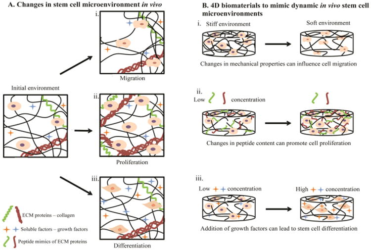
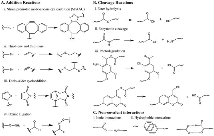
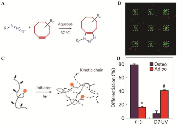
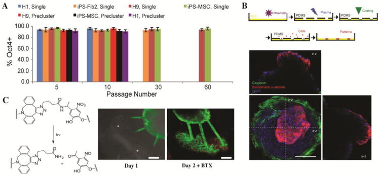
Stem cells reside in complex three-dimensional (3D) environments within the body that change with time, promoting various cellular functions and processes such as migration and differentiation. These complex changes in the surrounding environment dictate cell fate yet, until recently, have been challenging to mimic within cell culture systems. Hydrogel-based biomaterials are well suited to mimic aspects of these in vivo environments, owing to their high water content, soft tissue-like elasticity, and often-tunable biochemical content. Further, hydrogels can be engineered to achieve changes in matrix properties over time to better mimic dynamic native microenvironments for probing and directing stem cell function and fate. This review will focus on techniques to form hydrogel-based biomaterials and modify their properties in time during cell culture using select addition reactions, cleavage reactions, or non-covalent interactions. Recent applications of these techniques for the culture of stem cells in four dimensions (i.e., in three dimensions with changes over time) also will be discussed for studying essential stem cell processes.

Keywords: 3D cell culture; biomaterials; click chemistry; hydrogels; stem cells.


Figures

Figure 1
Figure 1. Changes in stem cell microenvironments in vivo captured by 4D biomaterials in vitro
A) The microenvironment of stem cells in the body changes with time. These changes have been observed to modulate cellular fate and functions, such as i) migration in response to gradients of matrix density/stiffness, ii) proliferation in response to matrix remodeling, and iii) differentiation in response to soluble factors (e.g., growth factors). B) Creating materials that capture such changes aids in the study of how cells respond to microenvironment remodeling events, such as wound healing or disease progression, toward ultimately directing these processes. For example, 4D hydrogel-based biomaterials have been engineered to enable i) changes in the mechanical properties of synthetic matrices by the addition or removal of crosslinks, influencing cell migration throughout the materials. At higher crosslink densities and moduli, cells have been entrapped within hydrogel-based matrices (left), whereas at lower crosslink densities and moduli cell migration has been observed (right). ii) Variation in biochemical content within the hydrogels through addition or removal of biochemical moieties (e.g., integrin-binding peptides or protein fragments) has been observed to promote cell proliferation. iii) Addition or sequestration of growth factors swollen within or tethered to the synthetic matrix has been observed to regulate stem cell differentiation. Note, these examples are meant to be representative, rather than comprehensive, of the ways 4D biomaterials have and can be used to probe stem cell processes; many extracellular cues influence multiple cellular functions (e.g., growth factors can influence migration, proliferation, and differentiation).
Figure 2
Figure 2. Chemistries to form and modify hydrogels for 4D culture of stem cells
A) ‘Click’ reactions, which occur under mild conditions and proceed close to 100% conversion, often have been used to form and modify hydrogels-based biomaterials in the presence of stem cells. These reactions have been used individually or in combination with each other. Here, a representative sample of the different functional groups and their reactions that have been used for hydrogel formation or modification in the presence of cells are noted: i) SPAAC, ii) thiol–ene and thiol– yne, iii) Diels-Alder cycloaddition, and iv) oxime ligation. B) Further, degradation or cleavage reactions have been used to change the properties of hydrogels during culture through the removal of crosslinks or pendant groups with i) preprogrammed hydrolysis, ii) cell-driven enzymatic hydrolysis, or ii) externally-triggered photolysis. C) Non-covalent interactions also have been used to form assembled hydrogels for use as dynamic stem cell culture matrices: i) ionic interactions, ii) hydrophobicity, and iii) hydrogen bonding.
Figure 3
Figure 3. 4D biomaterials for MSC culture
A) Photoreversible patterning of the protein vitronectin within well-defined hydrogel-based matrices controls encapsulated MSC differentiation. PEG-based hydrogels were formed by strain promoted azide-alkyne cycloaddition and B) patterned with vitronectin (dashed line regions), which promoted osteogenic differentiation (green stain indicates cells expressing the osteogenic marker osteocalcin). The regions of the hydrogel with no vitronectin exhibit limited MSC interaction with the matrix (e.g., rounded cell morphology) and limited osteogenic differentiation (no green), where all of the MSCs were stained with CellTracker Red. Adapted from DeForest and Tirrell with permission from Nature Publishing Group [62]. C) MMP-degradable HA hydrogels that allow MSC spreading and traction force generation (-) promote osteogenic differentiation. After secondary crosslinking at day 7 (D7 UV), spread cells are unable to degrade the matrix, affecting their ability to generate traction forces, and D) promoting differentiation into adipocytes instead of osteoblasts. Adapted from Burdick and coworkers with permission from Nature Publishing Group [31].
Figure 4
Figure 4. Stem and progenitor cell processes in 4D biomaterials
A) Long term expansion of hPSCs has been achieved in thermoreversible PNIPPAAm-PEG hydrogels. Seven hPSC lines were cultured for multiple passages within these materials with ∼ 95% pluripotency, indicated by Oct4 expression. The longest hPSC line passage accumulated to ∼ 1072 -fold expansion at 60 passages or 280 days. Adapted from Lei and Schaffer with permission from Proceedings of the National Academy of Sciences USA PNAS [57]. B) 3D cardiac microchamber was formed by confinement of iPSCs within PEG microwells. Cell-laden PEG microwells (top) were fabricated by PEG polymerization, PDMS mask and etching of the PEG film, coating of wells, and seeding of cells. Cells (bottom, nuclei blue) in the center differentiated into cardiomyocytes, as indicated by sarcomeric α-actinin stain (red). Cells along the perimeter differentiated into myofibroblasts, as indicated by calponin stain (green). x-z and y-z cross section projections (above and to the left) show the inner void space, indicating a 3D cardiac microchamber. Adapted from Healy and coworkers with permission from Nature Communications Publishing Group [77]. C) EB ESMNs were co-encapsulated with C2C12 cells within a photodegradable hydrogel. Channels (10 μm ×10 μm) were degraded between these two cell types using a 740-nm two-photon laser to cleave the o-nitrobenzyl-containing crosslinker (left). At two days, synapses (stained with alpha-bungarotoxin, red) were observed between motor axon extensions (green) and myotubes (right). Adapted from Anseth and coworkers with permission from Biomacromolecules the American Chemical SocietyACS Publications [37].

References

    1. National Institues of Health USD of H and HS. Stem Cell Basics. [accessed August 4, 2015];Stem Cell Inf. 2009 http://stemcells.nih.gov/staticresources/info/basics/SCprimer2009.pdf.
    1. Gattazzo F, Urciuolo A, Bonaldo P. Extracellular matrix: A dynamic microenvironment for stem cell niche. Biochim Biophys Acta. 2014;1840:2506–19. doi: 10.1016/j.bbagen.2014.01.010. - DOI - PMC - PubMed
    1. Rho JY, Kuhn-Spearing L, Zioupos P. Mechanical properties and the hierarchical structure of bone. Med Eng Phys. 1998;20:92–102. doi: 10.1016/S1350-4533(98)00007-1. - DOI - PubMed
    1. Watt FM, Huck WTS. Role of the extracellular matrix in regulating stem cell fate. Nat Rev Mol Cell Biol. 2013;14:467–73. doi: 10.1038/nrm3620. - DOI - PubMed
    1. Rao BM, Zandstra PW. Culture development for human embryonic stem cell propagation: Molecular aspects and challenges. Curr Opin Biotechnol. 2005;16:568–76. doi: 10.1016/j.copbio.2005.08.001. - DOI - PubMed