Review
. 2017 Jul 3:8:317.
doi: 10.3389/fneur.2017.00317. eCollection 2017.

Blood Pressure and Penumbral Sustenance in Stroke from Large Vessel Occlusion

Affiliations
Review

Blood Pressure and Penumbral Sustenance in Stroke from Large Vessel Occlusion

Robert W Regenhardt et al. Front Neurol. .

Abstract

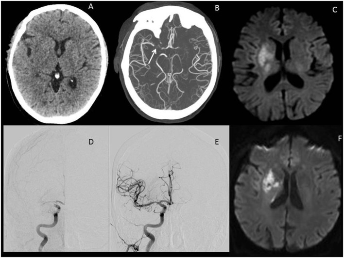
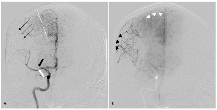
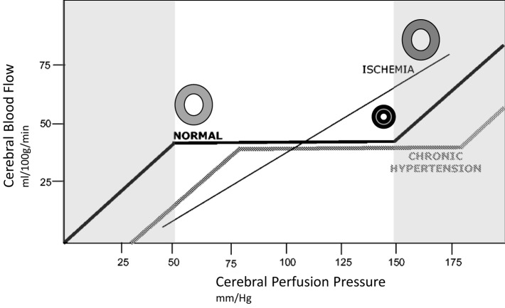
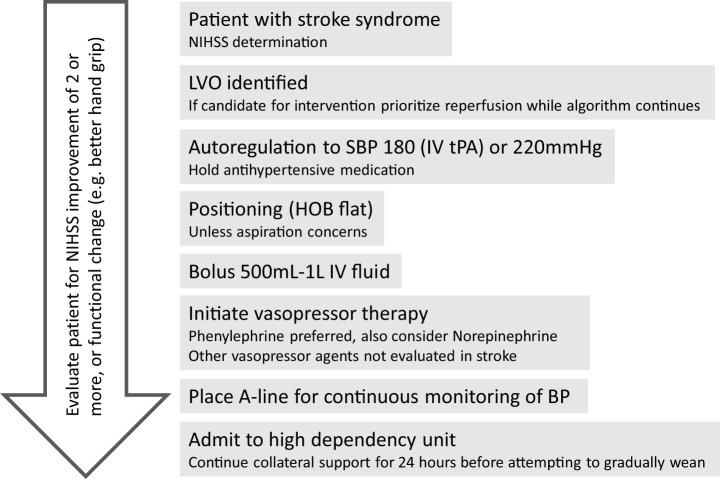
The global burden of stroke remains high, and of the various subtypes of stroke, large vessel occlusions (LVOs) account for the largest proportion of stroke-related death and disability. Several randomized controlled trials in 2015 changed the landscape of stroke care worldwide, with endovascular thrombectomy (ET) now the standard of care for all eligible patients. With the proven success of this therapy, there is a renewed focus on penumbral sustenance. In this review, we describe the ischemic penumbra, collateral circulation, autoregulation, and imaging assessment of the penumbra. Blood pressure goals in acute stroke remain controversial, and we review the current data and suggest an approach for induced hypertension in the acute treatment of patients with LVOs. Finally, in addition to reperfusion and enhanced perfusion, efforts focused on developing therapeutic targets that afford neuroprotection and augment neural repair will gain increasing importance. ET has revolutionized stroke care, and future emphasis will be placed on promoting penumbral sustenance, which will increase patient eligibility for this highly effective therapy and reduce overall stroke-related death and disability.

Keywords: blood pressure; neuroprotection; penumbra; pressor therapy; stroke; thrombectomy.


Figures

Figure 1
Figure 1
Illustrative images of a large vessel occlusion (LVO) stroke patient. Patient was a 67-year-old male presenting 4 h after onset with a full right middle cerebral artery (MCA) syndrome due to right MCA occlusion, NIHSS 14. (A) Emergent head computerized tomography without hemorrhage as a cause of stroke syndrome. (B) Axial maximal intensity projections from CTA showing right MCA occlusion (white arrow). (C) Emergent MRI DWI showing a small established core infarct. On the basis of this combined imaging and clinical data, it was determined that the patient had a large penumbra and small region of established injury and was therefore a good candidate for reperfusion therapy. (D) Anteroposterior view, catheter angiogram. The right internal carotid artery (ICA) injection reveals thrombus at the carotid terminus with only minimal anterior cerebral artery (ACA) opacification seen. Findings are consistent with an ICA-T occlusion. (E) Complete recanalization following mechanical thrombectomy, with full reperfusion (not shown) of the threatened penumbra. (F) 24 h MRI DWI showing arrest of infarct growth following reperfusion of the penumbra. The patient improved to NIHSS 4 by discharge on day 3 post-op. His stroke was determined to be cardioembolic following detection of atrial fibrillation after complete evaluation for cause, and he was free of deficits at 90-day follow-up.
Figure 2
Figure 2
AP angiographic images demonstrating adaptations of poorly perfused cerebral tissue, right internal carotid injection. Right middle cerebral artery (MCA) stroke, demonstrating poorly perfused but viable penumbral tissue as a therapeutic target through elevation of blood pressure. (A) Mid arterial phase. Black arrow: complete occlusion of MCA superior division. White arrow: partial occlusion of MCA inferior division. Dotted line: watershed zone between anterior and middle cerebral arteries. Small caliber vessels in this region represent leptomeningeal collaterals. Note early retrograde filling of distal superior division MCA territory (dotted arrow). (B) Late arterial phase. Black arrow heads demonstrate delayed antegrade fill of the inferior division and delayed retrograde fill of the superior division MCA branches. Note the maximal dilatation of these vessels, and the delay to parenchymal opacification when compared to the normally perfused anterior cerebral artery territory (white arrow heads). Both the partial occlusion with antegrade flow and the complete occlusion with retrograde collateral support benefit from systemic hypertensive responses, given the pressure passivity of the dilated distal MCA vasculature.
Figure 3
Figure 3
Idealized relationship between cerebral perfusion pressure (CPP) and cerebral blood flow (CBF). The normal autoregulation curve maintains a constant CBF over a range of CPP. At the lower end of the autoregulation range, vessels are dilated to encourage flow, at the upper end of the range they are maximally constricted, represented by circles in the illustration. Chronic hypertension moves the entire curve to the right. In ischemic brain, vasculature is maximally dilated, and the ability to autoregulate is lost. This introduces pressure passivity, where changes in CPP (through blood pressure modulation) are directly transmitted to CBF. The CBF is therefore passively dependent on the mean arterial pressure.
Figure 4
Figure 4
Algorithm for approach to hypertensive support for large vessel occlusion (LVO) patient. Reperfusion therapy should be the priority, but the algorithm provides for collateral support either while awaiting reperfusion (e.g., transfer to endovascular capable facility) or if reperfusion is not an option.

References

    1. Murray CJ, Lopez AD. Measuring the global burden of disease. N Engl J Med (2013) 369:448–57.10.1056/NEJMra1201534 - DOI - PubMed
    1. Lozano R, Naghavi M, Foreman K, Lim S, Shibuya K, Aboyans V, et al. Global and regional mortality from 235 causes of death for 20 age groups in 1990 and 2010: a systematic analysis for the Global Burden of Disease Study 2010. Lancet (2012) 380:2095–128.10.1016/S0140-6736(12)61728-0 - DOI - PMC - PubMed
    1. Goldstein LB, Adams R, Alberts MJ, Appel LJ, Brass LM, Bushnell CD, et al. Primary prevention of ischemic stroke: a guideline from the American Heart Association/American Stroke Association Stroke Council: cosponsored by the Atherosclerotic Peripheral Vascular Disease Interdisciplinary Working Group; Cardiovascular Nursing Council; Clinical Cardiology Council; Nutrition, Physical Activity, and Metabolism Council; and the Quality of Care and Outcomes Research Interdisciplinary Working Group: the American Academy of Neurology affirms the value of this guideline. Stroke (2006) 37:1583–633.10.1161/01.STR.0000223048.70103.F1 - DOI - PubMed
    1. Fisher CM. Lacunar strokes and infarcts: a review. Neurology (1982) 32:871–6.10.1212/WNL.32.8.871 - DOI - PubMed
    1. Lima FO, Furie KL, Silva GS, Lev MH, Camargo EC, Singhal AB, et al. Prognosis of untreated strokes due to anterior circulation proximal intracranial arterial occlusions detected by use of computed tomography angiography. JAMA Neurol (2014) 71:151–7.10.1001/jamaneurol.2013.5007 - DOI - PubMed