. 2015 Sep 1;6(9):5235-5245.
doi: 10.1039/c5sc01351h. Epub 2015 Jun 22.

An inhibitor of ubiquitin conjugation and aggresome formation

Affiliations

An inhibitor of ubiquitin conjugation and aggresome formation

Heeseon An et al. Chem Sci. .

Abstract

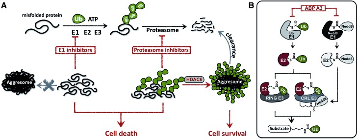
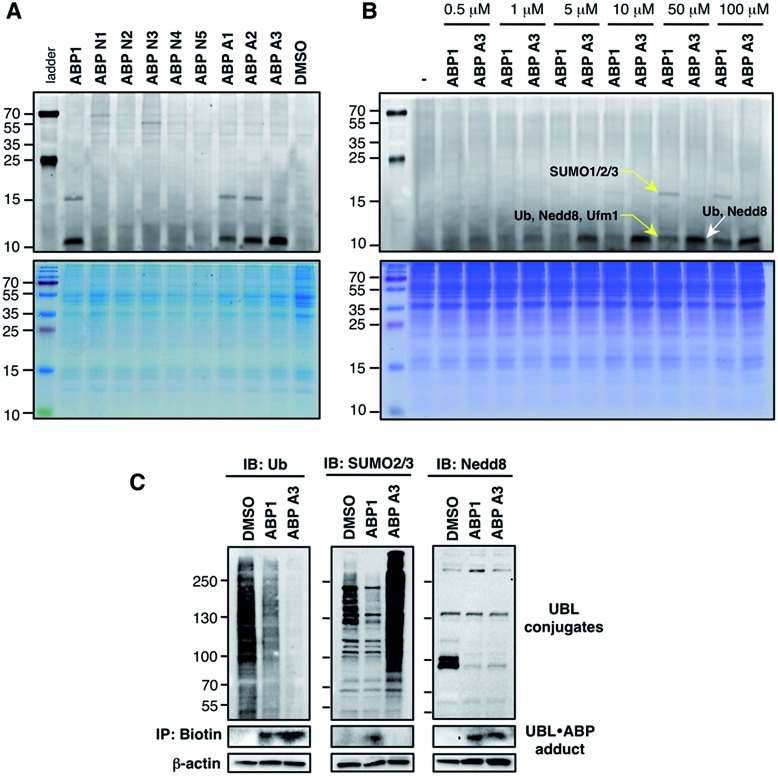
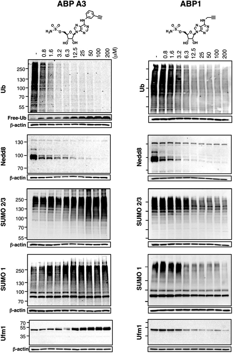
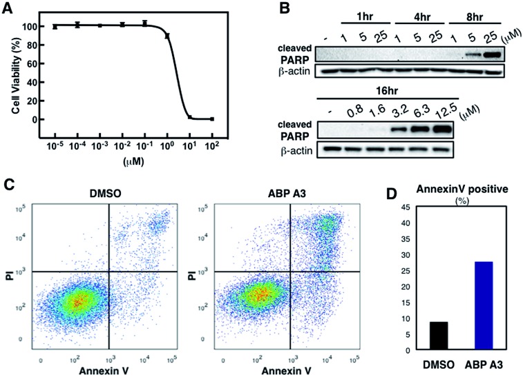
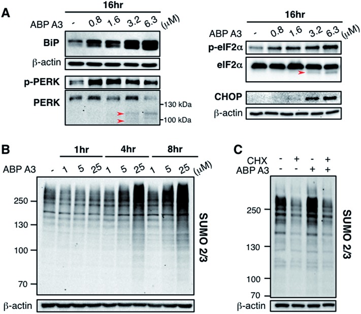
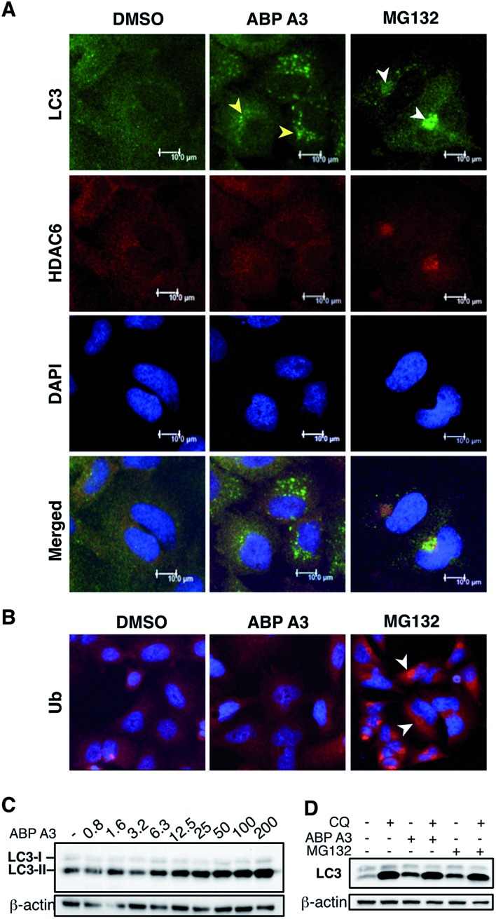
Proteasome inhibitors have revolutionized the treatment of multiple myeloma, and validated the therapeutic potential of the ubiquitin proteasome system (UPS). It is believed that in part, proteasome inhibitors elicit their therapeutic effect by inhibiting the degradation of misfolded proteins, which is proteotoxic and causes cell death. In spite of these successes, proteasome inhibitors are not effective against solid tumors, thus necessitating the need to explore alternative approaches. Furthermore, proteasome inhibitors lead to the formation of aggresomes that clear misfolded proteins via the autophagy-lysosome degradation pathway. Importantly, aggresome formation depends on the presence of polyubiquitin tags on misfolded proteins. We therefore hypothesized that inhibitors of ubiquitin conjugation should inhibit both degradation of misfolded proteins, and ubiquitin dependent aggresome formation, thus outlining the path forward toward more effective anticancer therapeutics. To explore the therapeutic potential of targeting the UPS to treat solid cancers, we have developed an inhibitor of ubiquitin conjugation (ABP A3) that targets ubiquitin and Nedd8 E1 enzymes, enzymes that are required to maintain the activity of the entire ubiquitin system. We have shown that ABP A3 inhibits conjugation of ubiquitin to intracellular proteins and prevents the formation of cytoprotective aggresomes in A549 lung cancer cells. Furthermore, ABP A3 induces activation of the unfolded protein response and apoptosis. Thus, similar to proteasome inhibitors MG132, bortezomib, and carfilzomib, ABP A3 can serve as a novel probe to explore the therapeutic potential of the UPS in solid and hematological malignancies.


Figures

Fig. 1
Fig. 1. Cellular effects of E1 enzyme inhibitors vs. proteasome inhibitors. (A) A fraction of newly synthesized proteins misfold, followed by their polyubiquitination and proteasomal degradation. Thus, proteasome inhibitors cause the accumulation of the polyubiquitinated misfolded proteins, which induces ER stress and contributes to cell death. However, misfolded polyubiquitinated proteins can be cleared by an alternative degradation pathway, which requires ubiquitin tags on misfolded proteins and involves the formation of aggresomes. In contrast to proteasome inhibitors, E1 enzyme inhibitors would induce the accumulation of misfolded proteins, yet would not cause the formation of aggresomes due to the lack of ubiquitin tags on the misfolded proteins. (B) Dual inhibitors of ubiquitin and Nedd8 E1 enzymes inhibit ubiquitin conjugation. Ubiquitin is activated by E1 enzyme, transferred onto the catalytic cysteine of E2, and conjugated to the lysine of protein substrates in the presence of RING or Cullin-RING E3s (CRL E3s). Alternatively E2–Ub thioesters can transfer ubiquitin onto the catalytic cysteine of HECT or RBR E3s (not shown), which then conjugate the ubiquitin onto the lysine of protein substrates. CRL E3s require the covalent modification with Nedd8 to be activated. Therefore, dual inhibition of ubiquitin E1 and Nedd8 E1 would efficiently inhibit substrate ubiquitination.
Fig. 2
Fig. 2. Mechanistic considerations in the design of selective inhibitors of E1 enzymes. (A) Mechanism of ABP1 analogue-mediated E1 inhibition (B) Structures of proposed ABP1 analogues.
Fig. 3
Fig. 3. (A) Schematic description of the intrinsic reactivity test of the nucleophile analogues using pre-activated ubiquitin thioester Ub–MES. (B) Deconvoluted mass spectra of the Ub–MES (100 μM) and ABP (1 mM) reaction mixture, after 2 hours of incubation at r.t. Reaction products are labeled as follows (1) Ub–MES, (2) ubiquitin, the hydrolysis product of Ub–MES, (3) Ub·ABP. * Ub·ABP N2 formed disulfide bond with free ABP N2, which was reduced to Ub·ABP N2 in the presence of TCEP (Fig. S1†).
Fig. 4
Fig. 4. (A) Dose dependent formation of Ub·ABP covalent adducts in the presence of UBE1 (0.5 μM), ATP (50 μM) and Ub (50 μM), after 2 hours at r.t. (B) The labeling efficiency of ABP1, ABP N2 and ABP A3 was compared via the quantification of the Ub·ABP levels at different concentrations. Each data point is shown as a triplicate (C) The inhibition potency of protein ubiquitination by ABP1, ABP N2 and ABP A3 was plotted based on the fluorescence intensity ratio of ubiquitin-modified GFP-Sic60 to total GFP-Sic60.
Fig. 5
Fig. 5. Labeling of proteins by ABPs in live A549 cells (A) A549 cells were treated with each ABP (100 μM) for 1 hour, followed by cell lysis, conjugation to fluorescent dye via copper-mediated [3 + 2] cycloaddition reaction, and in-gel fluorescence scanning. (B) Selectivity comparison between ABP1 and ABP A3 at different concentrations. (C) Correlation between UBL·ABP covalent adduct formation and the inhibition of the corresponding UBL conjugation pathways.
Fig. 6
Fig. 6. Intracellular selectivity of ABP A3 on UBL conjugation. A549 cells were treated with different concentrations of ABP A3 or ABP1 for 16 hours, lysed, and immunoblotted using anti-Ub, Nedd8, SUMO1, SUMO2/3 and Ufm1 antibodies. ABP A3 inhibited protein ubiquitination and neddylation, while not inhibiting but rather activating SUMOylation and ufmylation. In contrast, ABP1 inhibited all tested UBL conjugation systems.
Fig. 7
Fig. 7. Effect of ABP A3 on cell viability and apoptosis (A) A549 cells were treated with different concentrations of ABP A3 for 48 hours. Cell viability was monitored using CellTiter-Glo and analyzed with Prism software. Each data point is an average of six replicates. (B) ABP A3 treatment of A549 cells induced PARP cleavage and apoptosis (C) Flow cytometry analysis of ABP A3 treated cells (D) Total percentage of Annexin V-positive cells was plotted based on the flow cytometry data in (C).
Fig. 8
Fig. 8. Effect of ABP A3 on protein quality control in A549 cells (A) A549 cells treated with ABP A3 for 16 hours were immunoblotted with antibodies for UPR markers BiP, PERK, eIF2α, and the apoptosis marker CHOP. (B) Immunoblotting with anti-SUMO2/3 antibody revealed dramatic accumulation of SUMO conjugates in ABP A3-treated A549 cells. (C) A549 cells were treated with or without cycloheximide (30 μg ml–1) for 1 hour followed by the treatment with ABP A3 (10 μM) for an additional 3 hours. Inhibition of protein synthesis by cycloheximide restored ABP A3-induced SUMO conjugation to its basal levels.
Fig. 9
Fig. 9. Effect of ABP A3 on the aggresome-autophagy system in A549 cells. (A) A549 cells were treated with DMSO (0.025%), ABP A3 (2.5 μM) or MG132 (5 μM) for 24 hours, and immunostained with anti-LC3 and HDAC6 antibodies, and DAPI. Subsequent confocal images demonstrated the appearance of small autophagic puncta in ABP A3 treated cells (yellow arrows) and large aggresomes in MG132 treated cells (white arrows). (B) Cells prepared as in (A) were immunostained with ubiquitin antibody. (C) Immunoblotting of A549 cells treated with ABP A3 for 16 hours using anti-LC3 antibody showed accumulation of the lipidated form of LC3 (LC3-II) (D) A549 cells were treated with ABP A3 (5 μM) or MG132 (5 μM) in the presence or absence of chloroquine (CQ, 10 μM), followed by immunoblotting with LC3 antibodies. Increased amount of LC3-II upon ABP A3 + CQ treatment suggests the active autophagosome turnover in ABP A3 and MG132 treated cells.

References

    1. Haglund K., Dikic I. J. Cell Sci. 2012;125:265–275. - PubMed
    1. Lu Z., Hunter T. Annu. Rev. Biochem. 2009;78:435–475. - PMC - PubMed
    1. Trotman L. C., Wang X., Alimonti A., Chen Z., Teruya-Feldstein J., Yang H., Pavletich N. P., Carver B. S., Cordon-Cardo C., Erdjument-Bromage H., Tempst P., Chi S. G., Kim H. J., Misteli T., Jiang X., Pandolfi P. P. Cell. 2007;128:141–156. - PMC - PubMed
    1. Lohrum M. A., Woods D. B., Ludwig R. L., Balint E., Vousden K. H. Mol. Cell. Biol. 2001;21:8521–8532. - PMC - PubMed
    1. Geng J., Klionsky D. J. EMBO Rep. 2008;9:859–864. - PMC - PubMed