. 2017:2017:7407136.
doi: 10.1155/2017/7407136. Epub 2017 Jun 22.

Fusion to Flaviviral Leader Peptide Targets HIV-1 Reverse Transcriptase for Secretion and Reduces Its Enzymatic Activity and Ability to Induce Oxidative Stress but Has No Major Effects on Its Immunogenic Performance in DNA-Immunized Mice

Affiliations

Fusion to Flaviviral Leader Peptide Targets HIV-1 Reverse Transcriptase for Secretion and Reduces Its Enzymatic Activity and Ability to Induce Oxidative Stress but Has No Major Effects on Its Immunogenic Performance in DNA-Immunized Mice

Anastasia Latanova et al. J Immunol Res. 2017.

Abstract

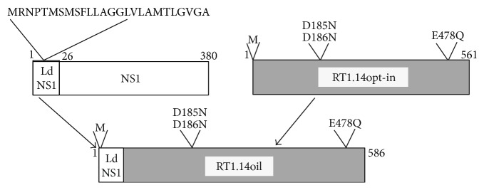
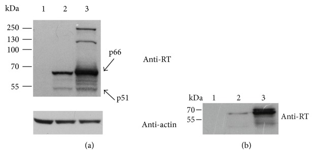
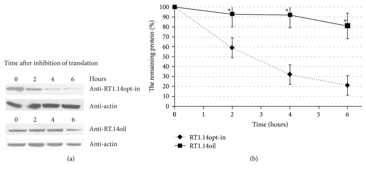
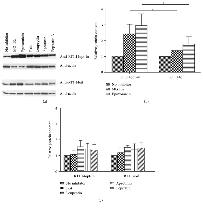
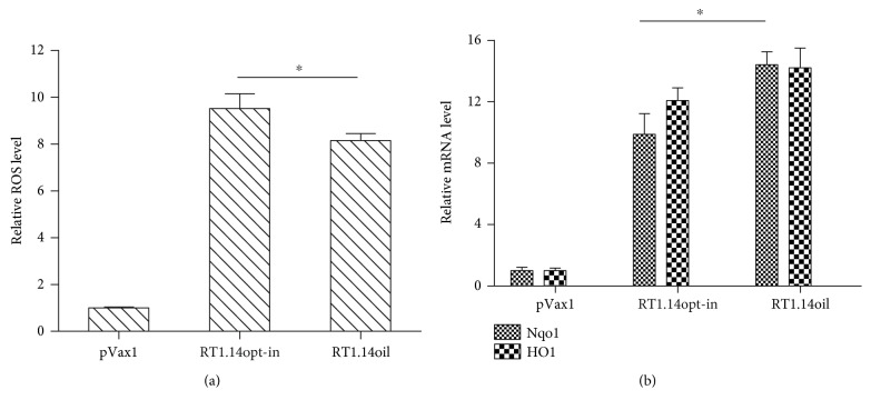
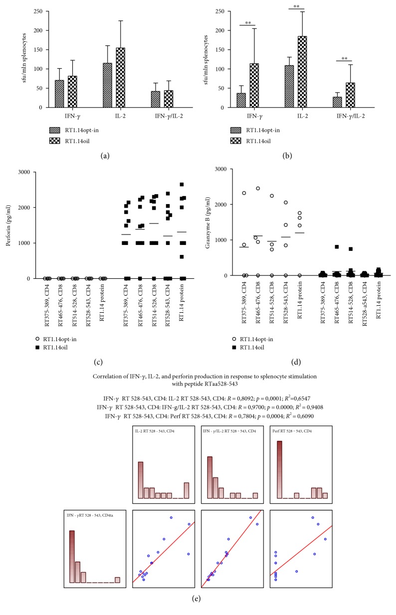
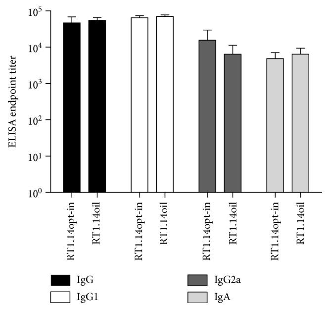
Reverse transcriptase (RT) is a key enzyme in viral replication and susceptibility to ART and a crucial target of immunotherapy against drug-resistant HIV-1. RT induces oxidative stress which undermines the attempts to make it immunogenic. We hypothesized that artificial secretion may reduce the stress and make RT more immunogenic. Inactivated multidrug-resistant RT (RT1.14opt-in) was N-terminally fused to the signal providing secretion of NS1 protein of TBEV (Ld) generating optimized inactivated Ld-carrying enzyme RT1.14oil. Promotion of secretion prohibited proteasomal degradation increasing the half-life and content of RT1.14oil in cells and cell culture medium, drastically reduced the residual polymerase activity, and downmodulated oxidative stress. BALB/c mice were DNA-immunized with RT1.14opt-in or parental RT1.14oil by intradermal injections with electroporation. Fluorospot and ELISA tests revealed that RT1.14opt-in and RT1.14oil induced IFN-γ/IL-2, RT1.14opt-in induced granzyme B, and RT1.14oil induced perforin production. Perforin secretion correlated with coproduction of IFN-γ and IL-2 (R = 0,97). Both DNA immunogens induced strong anti-RT antibody response. Ld peptide was not immunogenic. Thus, Ld-driven secretion inferred little change to RT performance in DNA immunization. Positive outcome was the abrogation of polymerase activity increasing safety of RT-based DNA vaccines. Identification of the molecular determinants of low cellular immunogenicity of RT requires further studies.


Figures

Figure 1
Figure 1
Schematic representation of chimeric drug-resistant HIV-1 reverse transcriptase carrying the N-terminal insertion of the N-terminal signal peptide of NS1 protein of TBEV (RT1.14oil). Rectangular boxes stand for polypeptide chains of NS1 with leader signal peptide (white) and RT1.14opt-in sequence (gray) with amino acid substitutions in polymerase (D185N, D186N) and RNase H signature motives (E478Q) leading to inactivation of respective enzymatic activities in the resultant RT1.14oil polyprotein. Amino acid positions in the polypeptide chain are designated with numbers.
Figure 2
Figure 2
Expression of HIV-1 RT1.14 fused to the N-terminal signal peptide of NS1 protein of TBEV in HeLa cells resolved by SDS-PAGE. (a) Western blotting of the lysates of HeLa cells transfected with pVax1 (1), pVaxRT1.14opt-in (2), and pVaxRT1.14oil (3) plasmids; upper panel, membranes stained with anti-RT; lower panel, with anti-β-actin antibodies for signal normalization. (b) Western blotting of the culture fluids of cells transfected with pVax1 (1), pVaxRT1.14opt-in (2), and pVaxRT1.14oil (3). Blots were stained with the specific polyclonal anti-RT antibodies as described in the experimental section “Materials and Methods.” To control equal loading of the samples of cell lysates on the gel, membranes were for the second time stained with anti-β-actin antibodies. Positions of the molecular mass markers are given to the left.
Figure 3
Figure 3
Cellular localization of RT without signal of secretion (RT1.14opt-in) and with a signal peptide (RT1.14oil) in HeLa cells. HeLa cells were transfected with the plasmids pVaxRT1.14opt-in (a, c, e) and pVaxRT1.14oil (b, d, f), grown on slides for 48 hours, fixed with methanol : acetone (1 : 1 v/v) (a, b, e, f) or 2% paraformaldehyde (c, d), and stained first with polyclonal anti-RT and with secondary FITC-conjugated anti-rabbit antibodies. For localization of RT in relation to ER, HeLa cells were treated with TRITC-conjugated antibodies to an ER marker calreticulin (e, f), and the overlay of FITC and TRITC signals is shown.
Figure 4
Figure 4
The kinetics of degradation of RT1.14opt-in and RT1.14oil in HeLa cells after blocking of translation with cycloheximide. (a) Western blotting of HeLa cells transfected with pVaxRT1.14opt-in and pVaxRT1.14oil plasmids 0, 2, 4, and 6 hours after cycloheximide treatment. The blots were stained with polyclonal anti-RT antibodies. To control equal loading of the samples on the gel, membranes were restained with anti-β-actin antibodies. (b) Diagram of protein degradation speed. 100% is according to initial amount of the protein. The graph is plotted in accordance with the results of 3 independent experiments, ±SD. p < 0,05 (Mann–Whitney test).
Figure 5
Figure 5
RT accumulation in HeLa cells treated with inhibitors of proteasomal and lysosomal proteolysis. Western blotting of HeLa cells transfected with pVaxRT1.14opt-in and pVaxRT1.14oil after 18-hour incubation with MG132 (5 μМ), epoxomicin (0,1 μМ), E64 (10 μМ), leupeptin (10 μg/ml), aprotinin (10 μg/ml), and pepstatin А (7,5 μМ) or without inhibitors. The blots were stained with monoclonal anti-RT antibodies. To control equal loading of the samples on the gel, the membranes were stripped and restained with anti-β-actin antibodies (a). Relative content of RТ1.14opt-in and RТ1.14oil in the samples treated with proteasomal inhibitors (b). Relative content of RТ1.14opt-in and RТ1.14oil in the samples treated with lysosomal inhibitors; no difference in response to lysosomal inhibitors between RT1.14opt-in- and RT1.14oil-expressing cells was detected (all p values > 0,05) (c). In (b) and (c), protein content in the untreated samples is taken for 1. Graphs in (b) and (c) represent the results of three independent experiments, +SD. p < 0,05 (Mann–Whitney test).
Figure 6
Figure 6
Induction of oxidative stress and oxidative stress response in cells expressing multidrug-resistant inactivated reverse transcriptase RT1.14opt-in and its derivative RT1.14oil carrying a signal peptide. Induction of the oxidative stress was detected as the production of ROS (a) and increase in the levels of mRNA of the phase II detoxification enzymes Nqo1 and HO1 (b). Levels of ROS were normalized to those in HEK293T cells transfected with the empty vector pVax1. Levels of mRNA for Nqo1 and HO-1 were normalized to levels of mRNA for actin and then represented as fold difference to the effect of empty vector pVax1. Data represent the results of two independent experiments, each done in triplicate, +SD. Results are compared using F test (Statistica AXA 10), p < 0,05.
Figure 7
Figure 7
Cellular immune response of mice immunized with pVaxRT1.14opt-in and pVaxRT1.14oil. IFN-γ, IL-2, and dual (IFN-γ/IL-2) secretion by splenocytes in response to stimulation with RT1.14 protein (a) or RT1.14-derived peptide aa528-543 (b) measured by Fluorospot. Responses represent the average number of signal-forming units (sfu) per mln cells in two independent experiment runs, each done in duplicate, +SD, n = 4, ∗∗p < 0,1 (Mann–Whitney test). Secretion of perforin (c) and granzyme B (d) by splenocytes of mice immunized with pVaxRT1.14opt-in and pVaxRT1.14oil in response to stimulation with RT1.14 protein and RT1.14-derived peptides. Splenocytes were stimulated with the recombinant RT 1.14 protein and RT peptides for 3 days. After that, cell culture fluids were collected and subjected to the analysis for granzyme B and perforin by sandwich ELISA. Data represent an average value of two repeated measurements for each mouse, in pg/ml. Correlation of the parameters of cellular immune response (e).
Figure 8
Figure 8
RT-specific IgGs and IgA induced in sera of mice immunized with pVaxRT1.14opt-in and pVaxRT1.14oil plasmids. Endpoint titers of RT-specific total IgG, IgG subtypes (IgG1, IgG2a), and IgA antibodies detected in the sera of BALB/c mice immunized with the genes encoding RT1.14opt-in and RT1.14oil plasmids. Data represent a mean + SD for the endpoint titer of antibodies against recombinant RT1.14 protein for four mice per group in two independent immunization runs. Cutoffs were set against the control mice immunized with empty vector pVax1 (see “Materials and Methods”). Groups demonstrate no significant difference in either IgG, or IgG1, or IgG2a, or IgA levels (p > 0,05).
Figure 9
Figure 9
Kinetics of in vivo bioluminescence from the sites of coadministration of RT genes and luciferase reporter genes. In vivo monitoring of luciferase activity on days 1, 3, 6, 9, 15, 22 after the administration of plasmids encoding RT1.14opt-in and RT1.14oil, or empty vector pVax1, each mixed with pVaxLuc encoding firefly luciferase (1 : 1 w/w). One curve represents bioluminescent emission from eight mice with two immunization sites in each, assessed in two independent immunization runs. p < 0,05 (Mann–Whitney test).

Similar articles

Cited by

References

    1. Jin X., Morgan C., Yu X., et al. Multiple factors affect immunogenicity of DNA plasmid HIV vaccines in human clinical trials. Vaccine. 2015;33(20):2347–2353. doi: 10.1016/j.vaccine.2015.03.036. - DOI - PMC - PubMed
    1. Munseri P. J., Kroidl A., Nilsson C., et al. Priming with a simplified intradermal HIV-1 DNA vaccine regimen followed by boosting with recombinant HIV-1 MVA vaccine is safe and immunogenic: a phase IIa randomized clinical trial. PLoS One. 2015;10(4, article e0119629) - PMC - PubMed
    1. Chen Y., Wang S., Lu S. DNA immunization for HIV vaccine development. Vaccine. 2014;2(1):138–159. doi: 10.3390/vaccines2010138. - DOI - PMC - PubMed
    1. Ahmed T., Borthwick N. J., Gilmour J., Hayes P., Dorrell L., Hanke T. Control of HIV-1 replication in vitro by vaccine-induced human CD8(+) T cells through conserved subdominant pol epitopes. Vaccine. 2016;34(9):1215–1224. doi: 10.1016/j.vaccine.2015.12.021. - DOI - PMC - PubMed
    1. Borthwick N., Ahmed T., Ondondo B., et al. Vaccine-elicited human T cells recognizing conserved protein regions inhibit HIV-1. Molecular Therapyy. 2014;22(2):464–475. doi: 10.1038/mt.2013.248. - DOI - PMC - PubMed

MeSH terms